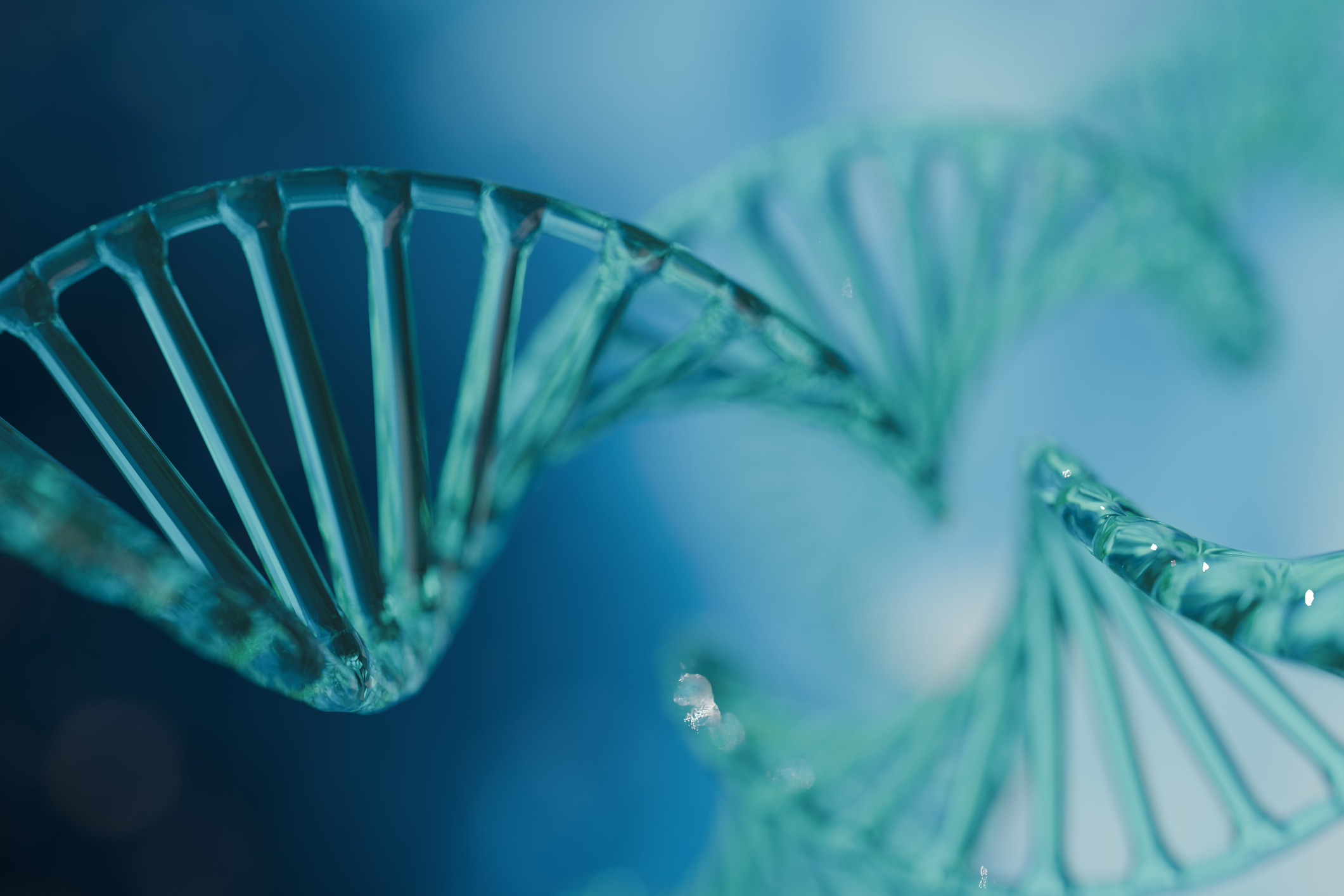
A recent study led by Paul DeCaen, PhD, associate professor of Pharmacology, has identified novel molecular mechanisms by which genetic mutations in the PKD2 gene cause the most common form of polycystic kidney disease, according to findings published in the Proceedings of the National Academy of Sciences.
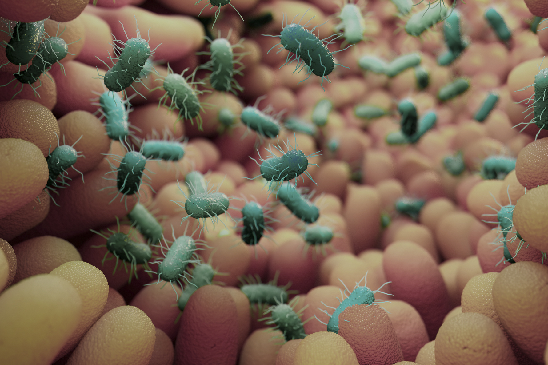
PKD2 encodes an ion channel localized to the primary cilia of cells lining the kidney collecting ducts, a series of tubules and ducts that helps achieve electrolyte and fluid balance in the body. Both inherited and acquired mutations in PKD2 are known to cause autosomal dominant polycystic kidney disease (ADPKD), a condition characterized by the growth of fluid-filled cysts in the kidneys that can lead to kidney failure and other serious complications.
According to the National Institute of Diabetes and Digestive and Kidney Diseases, one in 1,000 individuals will develop ADPKD and more than 95 percent of patients carry disease-causing genetic variants in PKD1 or PKD2. However, there are no available therapies that target these disease-causing variants.
“For over 20 years, we’ve known that certain genetic mutations cause this disease, yet we still lack a drug that can prevent cyst formation in affected individuals. As a first step toward therapeutic development, I set out to understand how these mutations alter the ion channel at the molecular level — both in terms of its structure and its function,” DeCaen said.
To better characterize these disease-causing genetic variants, DeCaen and his team used a combination of approaches, including direct cilia electrophysiology, cryogenic electron microscopy (cryo-EM) and super-resolution imaging, to study ADPKD cells expressing PKD2 mutations.
They discovered three germline missense variants — C632R, F629S and R638C — located in PKD2’s pore helix that cause defects in ion channel function, assembly and cilia trafficking, which helps transport proteins and other essential molecules to and from the cell.
“Even though all three mutations have impact in the same location of the channel, they have unique and unexpected effects,” DeCaen said.
While the F629S and R638C mutations impair PKD2’s ability to move ions efficiently in and out of the cilia, the C632R mutation misfolds the channel protein entirely, resulting in a complete loss of function, according to DeCaen.
The findings have the potential to inform the development of new targeted therapeutics that are tailored for each individual patient based on their specific genotype, DeCaen said.
“Our study suggests that a drug which corrects channel misfolding would be most effective for patients with the C632R mutation — this is called a molecular ‘corrector,’ a strategy which has been successfully used to treat conditions like cystic fibrosis. On the other hand, patients with the F629S or R638C mutations could be treated with therapeutics that reinstate channel function — this is called a channel ‘modulator,’ a strategy which has been successfully used to control cardiac arrhythmias and other ion channel diseases,” DeCaen said.
Orhi Esarte Palomero, PhD, a postdoctoral fellow in the DeCaen laboratory, was first author of the study. Eduardo Guadarrama, a student in the Driskill Graduate Program in Life Sciences (DGP), was a co-author of the study.
This work was supported by National Institutes of Health grant U24GM129547, the Ruth L. Kirschstein National Research Service Award individual postdoctoral fellowship (F32DK137477-01A1), NU Chicago KUH Forward training Grants (U2CDK129917 and TL1DK132769), the National Institute of Diabetes and Digestive and Kidney Diseases grants R01 DK123463-01 and R01 DK131118-01, and the PKD Foundation.