Feinberg
Northwestern Medicine | Northwestern University | Faculty Profiles

News Center

  • Categories
    • Campus News
    • Disease Discoveries
    • Clinical Breakthroughs
    • Education News
    • Scientific Advances
  • Press Release
  • Media Coverage
  • Podcasts
  • Editor’s Picks
    • COVID-19
    • Cardiology
    • Cancer
    • Neurology and Neuroscience
    • Aging and Longevity
    • Artificial Intelligence in Medicine
  • News Archives
  • About Us
    • Media Contact
    • Share Your News
    • News Feeds
    • Social Media
    • Contact Us
Menu
  • Categories
    • Campus News
    • Disease Discoveries
    • Clinical Breakthroughs
    • Education News
    • Scientific Advances
  • Press Release
  • Media Coverage
  • Podcasts
  • Editor’s Picks
    • COVID-19
    • Cardiology
    • Cancer
    • Neurology and Neuroscience
    • Aging and Longevity
    • Artificial Intelligence in Medicine
  • News Archives
  • About Us
    • Media Contact
    • Share Your News
    • News Feeds
    • Social Media
    • Contact Us
Home » New Technique Improves Proteoform Imaging in Human Tissue
Scientific Advances

New Technique Improves Proteoform Imaging in Human Tissue

By Melissa RohmanSep 9, 2022
Share
Facebook Twitter Email
Neil Kelleher, PhD, professor of Medicine in the Division of Hematology and Oncology, of Biochemistry and Molecular Genetics, and director of Northwestern’s Proteomics Center of Excellence, was senior author of the study published in Science Advances.

Investigators led by Neil Kelleher, PhD, professor of Medicine in the Division of Hematology and Oncology and of Biochemistry and Molecular Genetics, have developed a new imaging technique that increases the detection of intact proteoforms by fourfold when compared to current protein imaging methods.

The imaging technique, detailed in a recent paper published in Science Advances, provides high-resolution, high-throughput imaging of proteoforms, or all modified versions of proteins. Importantly, the technique is “label-free,” does not require antibodies and can identify whole proteoforms directly from any unfixed tissue. The technique can currently detect roughly 1,000 proteoforms and localizes proteoforms with a spatial resolution of 40 to 70 microns.

Several techniques are commonly used to image proteins in human tissue, but very few are capable of imaging proteoforms. Those that can image entire proteoforms do so by separating the proteoform from tissue and ionizing them for mass spectrometry. However, these techniques offer low molecular specificity.  

To address this issue, Kelleher’s team developed proteoform imaging mass spectrometry (PiMS). The technique works by extracting proteoforms from the tissue with nanodroplets, “weighing” the extracted proteoforms to identify them and then using this data to construct proteoform images of the scanned tissue.

“The real innovation with PiMS is that it couples a robust existing technique for extracting and ionizing proteoforms, nanoDESI, with a breakthrough technology for individual ion mass spectrometry that was co-invented by Thermo Fisher Scientific and Northwestern Proteomics. Compared to regular detection techniques, individual ion mass spectrometry offers up to 500 times more sensitivity and 20 times more resolving power. That significantly increases the power of the technique, and PiMS detects larger, rarer proteoforms and greatly extends the limits of proteome coverage,” said Kelleher, who is also director of Northwestern’s Proteomics Center of Excellence, the Robert H. Lurie Comprehensive Cancer Center’s Proteomics Core Facility and of Northwestern’s Chemistry of Life Processes Institute.

PiMS images of individual proteoforms that selectively illuminate different anatomical regions and cellular neighborhoods.

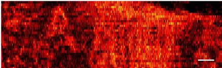
To demonstrate PiMS’ capabilities, Kelleher’s team used the technique to image proteoforms from functional units of the human kidney. These images revealed distinct spatial localizations of proteoforms from different anatomical regions and functional tissue units like the renal cortex versus the medulla.

PiMS’ increased proteome coverage also opens the door for wider applications in molecular tissue mapping, identifying novel biomarkers and improving disease diagnosis, according to Kelleher.

“Recently, there has been a big push in genomics and proteomics for single-cell biology: to better capture the heterogeneity of diseases by using spatial or single-cell approaches that preserve the many diverse signals instead of bulk approaches that mix all cell types and regions together. The spatial approach in particular adds a far greater precision for protein imaging and we are currently pushing it to identify thousands of proteoforms with single-cell resolution,” Kelleher said.

This work was supported by National Institutes of Health grants UH3 CA246635, P41 GM108569, P30 DA018310 and P30 CA060553.

Biochemistry and Molecular Genetics Cancer Medicine Research
Share. Facebook Twitter Email

Related Posts

Shortage of Mental Health Professionals Linked to Increase in Youth Suicides

Jan 25, 2023

Understanding How Childhood Brain Tumors Develop

Jan 23, 2023

Medical School Establishes New Center for Psychiatric Neuroscience

Jan 19, 2023

Comments are closed.

Latest News

Changes in Medical School Leadership

Jan 26, 2023

Shortage of Mental Health Professionals Linked to Increase in Youth Suicides

Jan 25, 2023

Northwestern Medicine Scholars Program Inspires a New Generation of Physicians and Scientists 

Jan 24, 2023

Understanding How Childhood Brain Tumors Develop

Jan 23, 2023

Medical School Establishes New Center for Psychiatric Neuroscience

Jan 19, 2023
  • News Center Home
  • Categories
  • Press Release
  • Media Coverage
  • Editor’s Picks
  • News Archives
  • About Us
Flickr Photos
Feinberg_In_Vivo_20221209_tcrawford-24
Feinberg_In_Vivo_20221209_tcrawford-16
Feinberg_In_Vivo_20221209_tcrawford-14
Feinberg_In_Vivo_20221209_tcrawford-5
Feinberg_In_Vivo_20221209_tcrawford-6
Feinberg_In_Vivo_20221209_tcrawford-10
Feinberg_In_Vivo_20221209_tcrawford-8
Feinberg_In_Vivo_20221209_tcrawford-18
Feinberg_In_Vivo_20221209_tcrawford
Feinberg_In_Vivo_20221209_tcrawford-23
Feinberg_In_Vivo_20221209_tcrawford-25
Feinberg_In_Vivo_20221209_tcrawford-26

Northwestern University logo

Northwestern University Feinberg School of Medicine

RSS Facebook Twitter LinkedIn Flickr YouTube Instagram
Copyright © 2023 Northwestern University
  • Contact Northwestern University
  • Disclaimer
  • Campus Emergency Information
  • Policy Statements

Type above and press Enter to search. Press Esc to cancel.