This story was originally published in the August 2025 issue of the Breakthroughs newsletter.
A new core facility is opening this month to provide Northwestern investigators with access and training to use a Glacios-2 Cryo-Transmission Electron Microscope (X-FEG 200 kV, Falcon 4i detector and Selectris energy filter). The Feinberg Advanced Cryo Electron-Microscopy and Tomography (FACET) core is located in the sub-basement of the Simpson Querrey Biomedical Research Center.

The new core will provide access to the Glacios-2 Cryo-Transmission Electron Microscope, ancillary equipment for grid preparation and expertise as a shared resource to enhance research throughout the University. Overall, the new facility will contribute directly to Northwestern’s mission to foster innovation and support investigations into the biological underpinnings of human disease.
This technology has been spreading across research universities since the Nobel Prize for Chemistry was awarded in 2017 for the development of cryo-EM for the high-resolution structure determination of biomolecules, which is testament to the impact of these methodologies. The technology was further developed and packaged by companies like Thermo Fisher Scientific to market this instrumentation.
Unlike a light microscope, which utilizes photons, this cryo-TEM microscope uses a beam of electrons to visualize vitrified protein samples, said Paul DeCaen, PhD, associate professor of Pharmacology.
While at Harvard in 2013, DeCaen learned of transformative technological advances in cryo-EM, such as direct electron detection, which helped democratize, and later broke the barriers for this approach. He joined Northwestern in 2016 and had his lab staff trained to use a cryo-EM instrumentation. He began to explore what it would take to bring the cryo-EM technology to Feinberg.
In an informal faculty “chalk talk” held by the Department of Pharmacology, DeCaen raised the importance of this technology and the benefits that it could bring to scientists on the medical campus. He was able to pitch the idea to Jeff Weiss, PhD, director for research core planning, and Rex Chisholm, PhD, vice dean for scientific affairs and graduate education, who explored options for funding. Through a strategic initiative from the dean’s office and a block grant from the State of Illinois, the new instrument was purchased and installed.
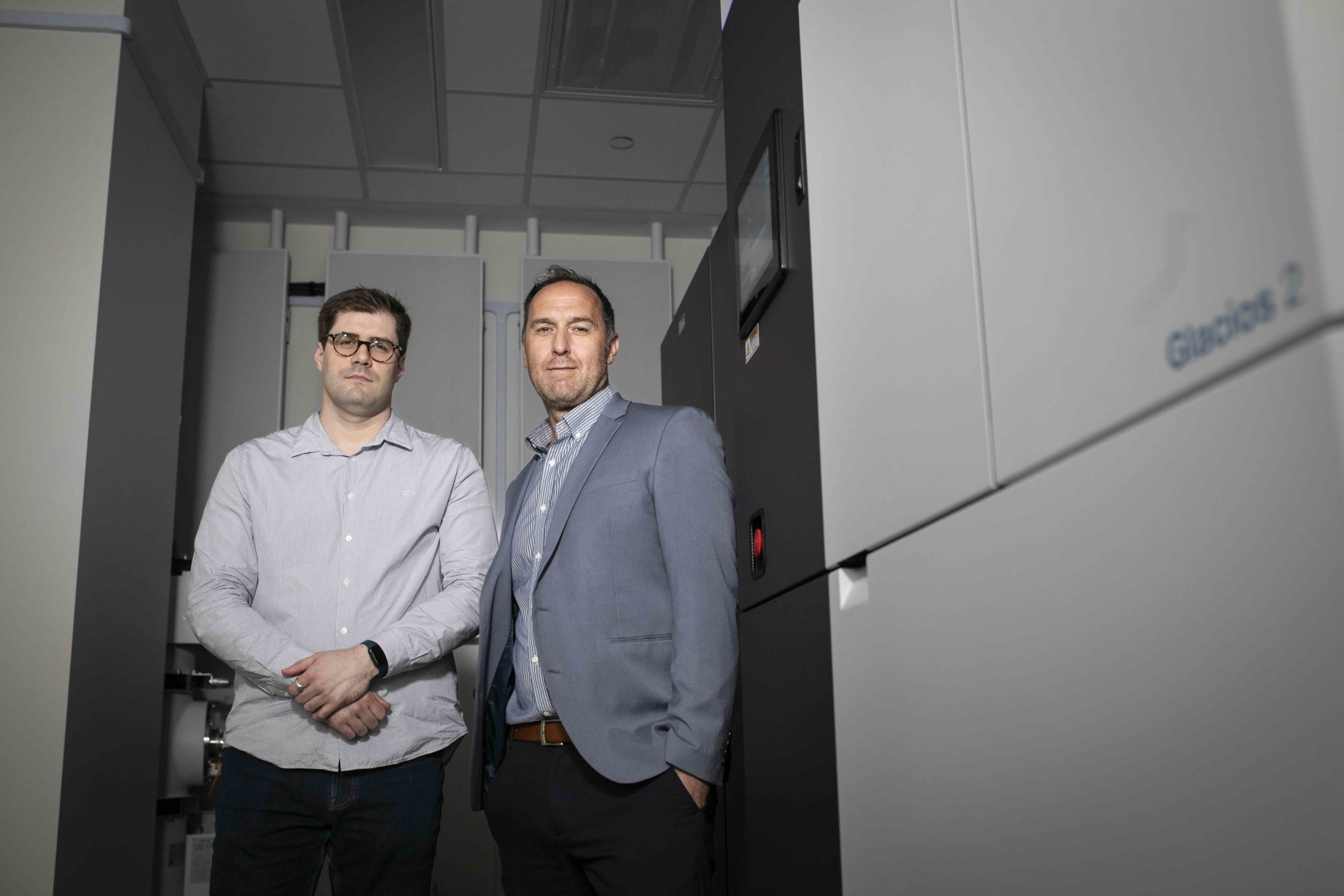
The new core will be led by Orhi Esarte Palomero, PhD. Previously a post-doctoral fellow in DeCaen’s lab, Esarte Palomero has trained extensively in the national cryo-EM centers at the Pacific Northwest Center for Cryo-EM (PNCC) and the Stanford-SLAC CryoEM Center.
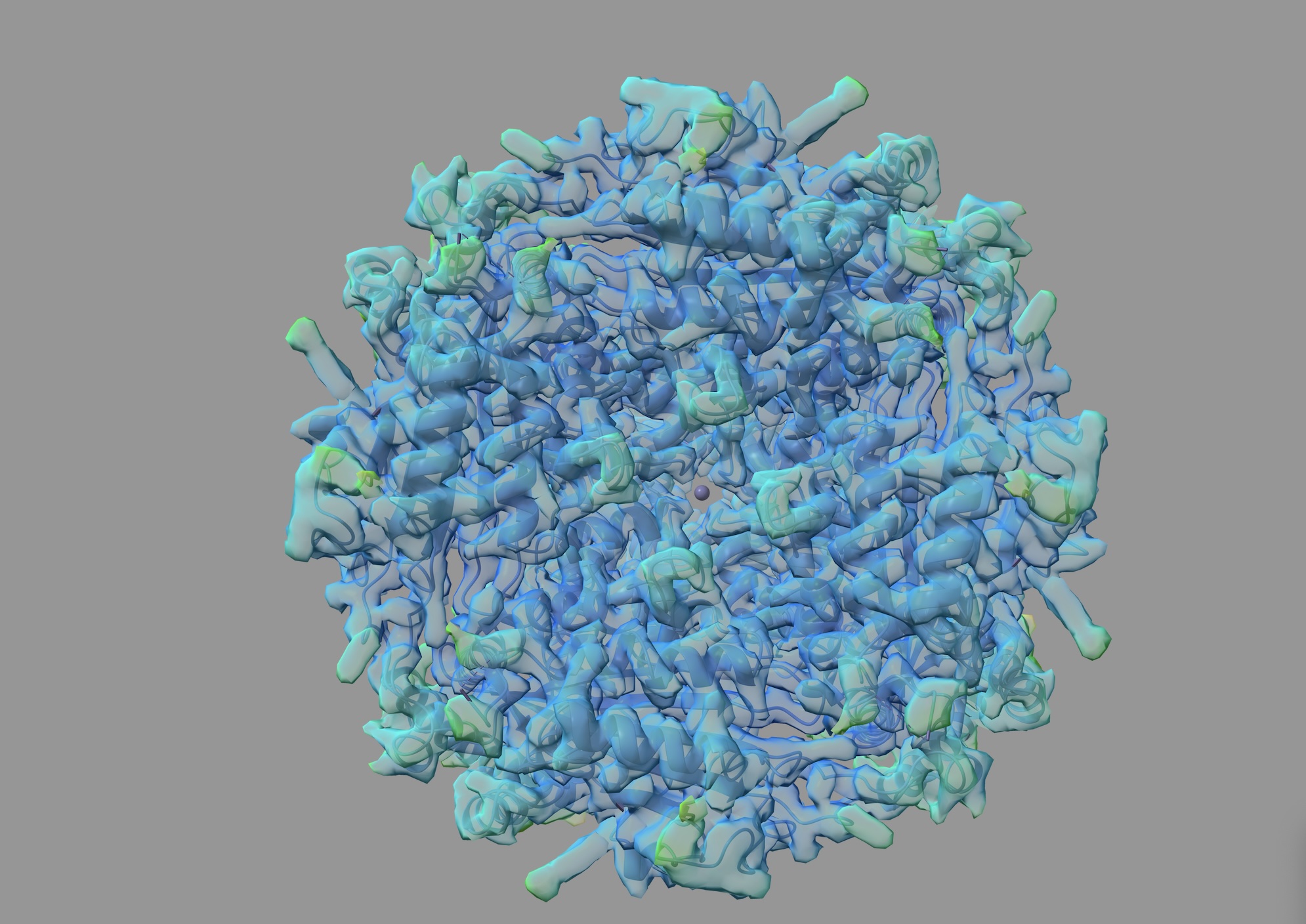
The Glacios uses electrons to image small biomolecules, which allow scientists to reconstruct the structure of these tiny molecules, said Esarte Palomero.
Decaen’s lab studies polycystic kidney disease caused by disruptive mutations in genes that encode an ion channel found in renal primary cilia. As these cells lose polarity, they start to form cysts in the kidney, which can lead to fatal kidney failure if the patient does not receive a transplant.
“The microscope will allow us to visualize the molecular chemical features of the channel at the atomic level. The resulting structural template will be used to develop prototypic drugs to treat polycystic kidney disease,” DeCaen said. “In the near future, we will also use this microscope to visualize why cilia organelles become unstable and stop being formed in patient cells. This cutting-edge approach is called cryo-tomography, and we will be able to see how proteins within cilia fail to assemble. These types of questions are at the at the forefront of this technology and our molecular understanding of human diseases.”
A recent study published in the Proceedings of the National Academy of Sciences led by DeCaen used cryo-EM technology to identify novel molecular mechanisms by which genetic mutations in the PKD2 gene cause the most common form of polycystic kidney disease. The findings have the potential to inform the development of new targeted therapeutics that are tailored for each individual patient based on their specific genotype, DeCaen said.
Investigators who may be interested in using the microscope could be anyone studying an organelle-level change, organelle structure, disease-associated organelles and/or protein structure. Anyone looking to characterize a structure in just about any cell would benefit from using the microscope.
“Structural biology and cryo-EM can advance our understanding of the pathologies that our [scientists] are working on. We hope that labs will incorporate the technology in an interdisciplinary way,” said Esarte Palomero.
According to DeCaen, while the Human Genome Project helped to enumerate genetic components, this new technology defines their structural protein products and their location within cells to elucidate how we approach therapeutics.
Esarte Palomero said he looks forward to welcoming users to the new core and providing training and assistance to get the most of this new asset.
“There are a lot of opportunities to understand disease mechanisms, patient variants, disease progression and even discover new therapies through the lens of cryo-EM. Now that we have state of the art tools to gain structural biology insights, it will enhance the quality, and the reach of the research work we do at Northwestern” said Esarte Palomero.
More information is available about the FACET core here.