Northwestern Medicine scientists have developed a new non-invasive approach that could help better determine which patients with glioblastoma are responding favorably to chemotherapy treatment and inform future treatment plans, according to a recent study published in Nature Communications.
Glioblastoma is the most common and aggressive primary brain tumor in adults, with a five-year survival rate of less than 7 percent, according to the National Brain Tumor Society. The cancer is notoriously difficult to treat because it is highly invasive and because the blood-brain barrier, which acts as a protective filter for the brain, prevents many chemotherapy drugs from reaching the tumors.
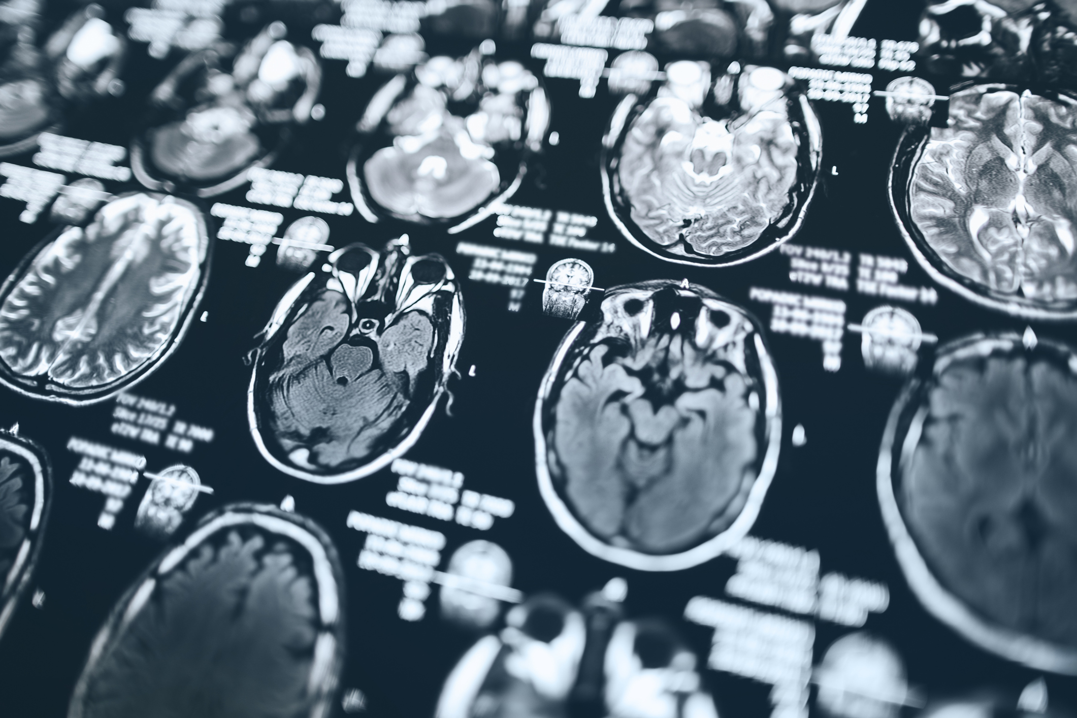
An additional challenge in glioblastoma is assessing therapeutic response, as radiation or immunotherapies can distort contrast-enhancing regions on MRI, leading to uncertainty about what constitutes true tumor progression versus treatment-associated ‘pseudo-progression’.
Determining whether a glioblastoma tumor is responding to therapy or continuing to grow may influence future management choices, such as the need for additional surgery, radiation or chemotherapy. This can also be important for determining the success of new therapeutic approaches in the context of a clinical trial. Unfortunately, performing month-to-month MRI scans of the brain may not always produce clear and accurate results, said Adam Sonabend, MD, associate professor of Neurological Surgery and senior author of the study.
“Ultimately, what we have to rely on is waiting long enough to see if the patient ultimately lives longer or shorter than expected. As you might imagine, that’s not practical for brain tumor patients when time is very valuable,” said Sonabend, who is also a member of the Robert H. Lurie Comprehensive Cancer Center of Northwestern University and a member of the Lou and Jean Malnati Brain Tumor Institute at the Lurie Cancer Center.
In a previous first-of-its-kind clinical trial, Sonabend’s team used a skull-implantable ultrasound device in patients with glioblastoma to open the blood-brain barrier and deliver chemotherapy that was injected intravenously.

Results from the phase I clinical trial showed that opening the blood-brain barrier using this device led to a four- to six-fold increase in chemotherapy drug concentrations of paclitaxel and carboplatin in the brain. Those drugs aren’t typically used to treat glioblastoma because they don’t cross the blood-brain under normal conditions.
Expanding upon these findings, in the current study Sonabend’s team used a non-invasive liquid biopsy approach — a blood test that detects cancer material circulating in the bloodstream — to determine which patients were responding well to paclitaxel chemotherapy versus those were not.
Blood samples were analyzed that had been collected longitudinally during chemotherapy treatment and blood-brain barrier opening in the phase I trial (every three weeks until disease progression or for up to six cycles).
The team collaborated with scientists from the University of Michigan to develop a microfluidic chip that could capture glioblastoma-associated exosomes based on the expression of phosphatidylserine, a phospholipid associated with cellular stress. Exosomes from the tumor microenvironment were detected abundantly in the blood of patients, and carry proteins, nucleic acids and other materials from their cell of origin.
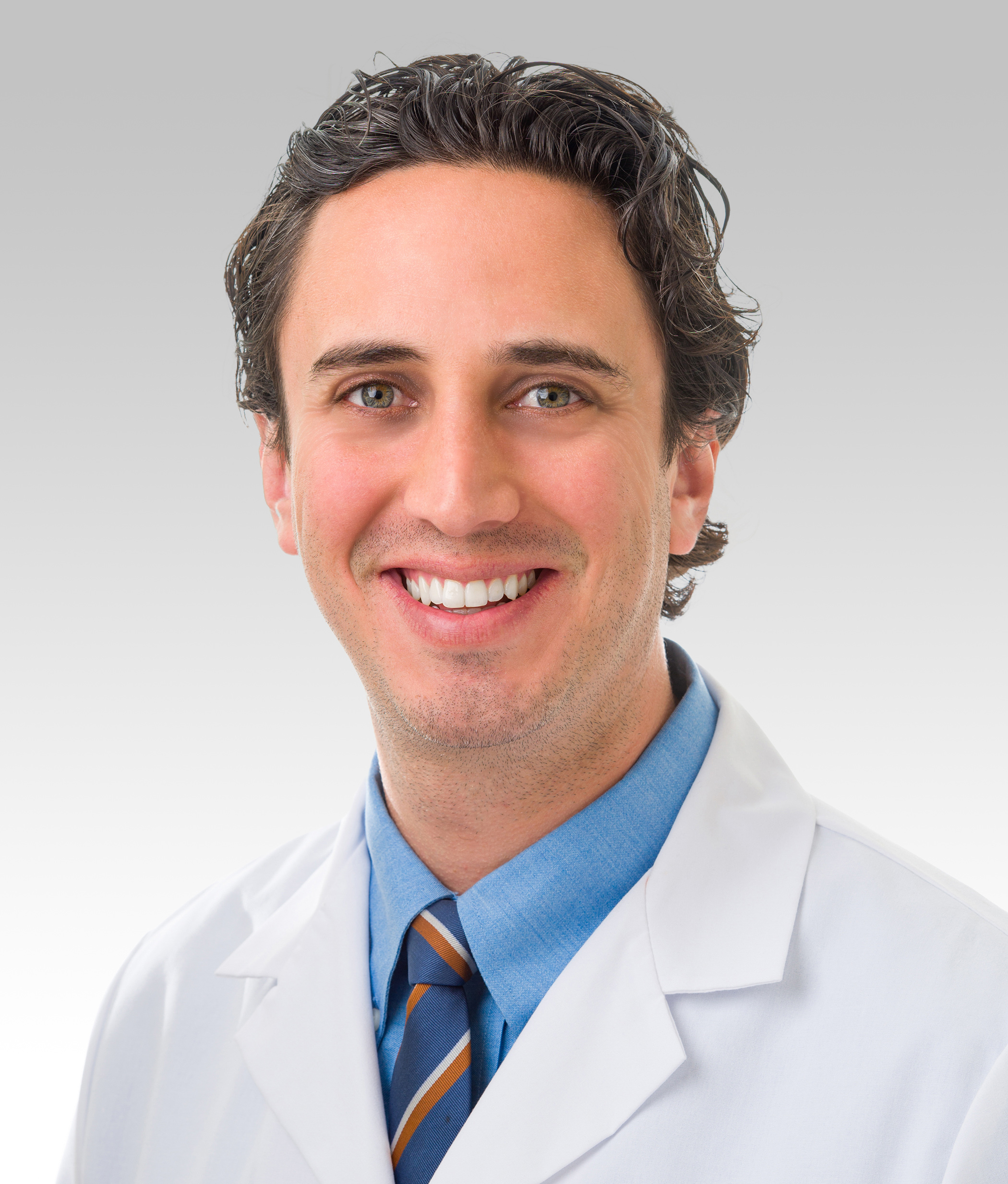
“This approach could be helpful for the clinical management of glioblastomas, because captured material gives a less-invasive and up-to-date snapshot of the tumor’s behavior. Previously, we needed to perform an additional surgery to determine this information,” said Mark Youngblood, MD, PhD, a resident in Neurological Surgery and first author of the study.
By capturing these circulating exosomes after each round of chemotherapy treatment, the scientists identified differences in the responses of exosomes in patients who would go on to respond favorably to chemotherapy versus those who did not.
They found that while paclitaxel-susceptible glioblastoma cell lines shed exosomes during therapy-related induction of apoptosis, or cell death, chemotherapy-resistant glioblastoma cell lines did not.
“We think this is driven by glioblastoma cells responding to chemotherapy and undergoing cell death, which results in shedding of more exosomes that can be detected using our microfluidic devices,” Youngblood said.
The findings suggest the approach could be used as a real-time assessment for predicting chemotherapy response after blood-brain barrier opening in glioblastoma patients, which can help better inform a patient’s course of treatment and ultimately improve outcomes.
“Our study introduces an efficient microfluidic platform for the capture of circulating glioblastoma extracellular vesicles and particles and demonstrates that release upon blood-brain barrier opening is predictive of outcomes following paclitaxel treatment. This approach represents a real-time surrogate biomarker for treatment response for a disease where imaging-based assessment of response has not been shown to be reliable,” the authors wrote.
Co-authors include Karl Habashy, MD, a resident physician in Neurological Surgery; Mateo Gomez, a research assistant in the Youngblood lab; Harrshavasan Congivaram, a third-year medical student; Hui Zhang, PhD, professor of Preventive Medicine in the Division of Biostatistics and Informatics; Katarzyna Pituch, PhD, research assistant professor of Neurological Surgery; Ditte Primdahl, PhD, assistant professor in the Ken and Ruth Davee Department of Neurology and of Neurological Surgery; Karan Dixit, MD, assistant professor of Neurology in the Divisions of Neuro-oncology and Hospital Neurology; Rimas Lukas, MD, vice chair of Outpatient Neurology in the Department of Neurology; Priya Kumthekar, MD, ’11, ‘12 GME, associate professor of Neurology in the Division of Neuro-oncology; Crismita Dmello, PhD, research assistant professor of Neurological Surgery; and Roger Stupp, MD, the Paul C. Bucy Professor of Neurological Surgery and chief of Neuro-oncology in the Department of Neurology.
Kumthekar, Dmello and Stupp are also members of the Lurie Cancer Center.
This work was supported by an American Brain Tumor Association Basic Research Fellowship (fully supported by Tap Cancer Out), National Institutes of Health 1R01CA245969-01A1, P50CA221747, 5-R33-CA-202867, 1-R01-CA-208335-01-A1, and 1U19CA264338-01; the Lou and Jean Malnati Brain Tumor Institute; the Moceri Family Foundation in memory of Sharon Moceri; and Tina and Vic Kedaitis.