Feinberg
Northwestern Medicine | Northwestern University | Faculty Profiles

News Center

  • Categories
    • Campus News
    • Disease Discoveries
    • Clinical Breakthroughs
    • Education News
    • Scientific Advances
    • Podcast
  • Press Release
  • Media Coverage
  • Editor’s Picks
    • COVID-19
    • Cardiology
    • Cancer
    • Neurology and Neuroscience
    • Aging and Longevity
    • Artificial Intelligence in Medicine
  • News Archives
  • About Us
    • Media Contact
    • Share Your News
    • News Feeds
    • Social Media
    • Contact Us
Menu
  • Categories
    • Campus News
    • Disease Discoveries
    • Clinical Breakthroughs
    • Education News
    • Scientific Advances
    • Podcast
  • Press Release
  • Media Coverage
  • Editor’s Picks
    • COVID-19
    • Cardiology
    • Cancer
    • Neurology and Neuroscience
    • Aging and Longevity
    • Artificial Intelligence in Medicine
  • News Archives
  • About Us
    • Media Contact
    • Share Your News
    • News Feeds
    • Social Media
    • Contact Us
Home » Cytoskeletal Proteins Interact to Form Intracellular Networks
Scientific Advances

Cytoskeletal Proteins Interact to Form Intracellular Networks

By Melissa RohmanApr 7, 2022
Share
Facebook Twitter Email
Robert Goldman, PhD, professor of Cell and Developmental Biology and of Medicine in the Division of Pulmonary Care, was a co-author of the study published in the published in Proceedings of the National Academy of Sciences.

Investigators have discovered that two cytoskeletal proteins which were previously thought to function independently actually interact and form cytoskeletal networks within the cell surface, according to findings published in Proceedings of the National Academy of Sciences (PNAS).

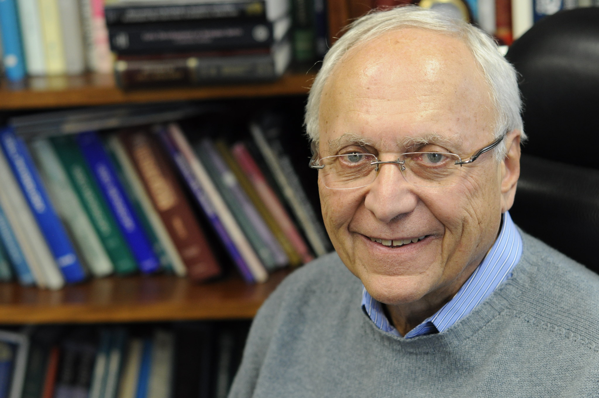
“These findings significantly alter the definition and function of the cell cortex,” said Robert Goldman, PhD, professor of Cell and Developmental Biology, of Medicine in the Division of Pulmonary Care and a co-author of the study.

The cytoskeleton of eukaryotic cells is composed of various networks comprising several types of filamentous proteins, including actin and intermediate filaments.

Notably, filamentous actin (F-actin) and vimentin intermediate filaments support cell structure and regulate numerous cell functions; F-actin is found in the cell cortex, closely associated with the surface membrane and regulates the cell’s contractility and movement, while the majority of vimentin intermediate filaments are located within the cell and regulate its shape, structure and mechanical properties during cell movement and migration.

These cytoskeletal proteins have previously been thought to form separate intracellular networks and function independently of one another. However, the current study revealed that wasn’t the case.

Using advanced cell imaging to study cell cultures expressing F-actin and vimentin, the investigators discovered that these cytoskeletal proteins actually work synergistically in the cell cortex.

Fluorescence image of a MEF cell protrusion. Emerald-vimentin is shown in green. The white square indicates the position where a cryo-tomogram is acquired.

They found that this interaction not only helps regulate the cell’s contractility, but also that vimentin intermediate filaments help regulate actin polymerization, promoting proper cell signaling.

“The findings are very basic but certainly reflect on many aspects of cell physiology and cell migration within developing tissue and organ systems as well as mechanisms responsible for normal cell movements within complex tissues,” said Goldman, who is also a member of the Robert H. Lurie Comprehensive Cancer Center of Northwestern University.

Goldman added that the findings could also have pathological implications, such as in the case of wound healing and the regulation of cell movement in cancer metastasis, both of which require proper function of a cell’s mechanical properties — supported by vimentin intermediate filaments — and contractility — enabled by F-actin and its associated proteins.

Stephen Adam, ’86 PhD, associate professor of Cell and Developmental Biology, and Suganya Sivagurunathan, PhD, a postdoctoral fellow in the Goldman laboratory, were co-authors of the study. Collaborators included David Weitz, PhD, the Mallingkrodt Professor of Physics and Applied Physics at Harvard University, and Ohad Medalia, PhD, professor of biochemistry at the University of Zurich.

This work was supported by a National Institutes of Health Program Project grant (2P01GM096971), which has been organized and directed by Goldman for almost a decade.

Cell and Developmental Biology Research
Share. Facebook Twitter Email

Related Posts

Hospitals Bound to Patient Safety Rules that Aren’t all Backed by Evidence

Jun 24, 2022

Identifying Protein Interactions that Promote Cancer Growth

Jun 24, 2022

Calcium Channel Blockers May Improve Chemotherapy Response

Jun 21, 2022

Comments are closed.

Latest News

Student Spearheads Ukraine Aid Efforts

Jun 27, 2022

Hospitals Bound to Patient Safety Rules that Aren’t all Backed by Evidence

Jun 24, 2022

Identifying Protein Interactions that Promote Cancer Growth

Jun 24, 2022

Combination Treatment May Improve Quality of Life in Kidney Cancer

Jun 23, 2022

Calcium Channel Blockers May Improve Chemotherapy Response

Jun 21, 2022
  • News Center Home
  • Categories
  • Press Release
  • Media Coverage
  • Editor’s Picks
  • News Archives
  • About Us
Flickr Photos
pride7
pride6
pride5
pride4
pride3
pride2
pride1
20220607_Feinberg Campus_0070
20220607_Feinberg Campus_0066
20220607_Feinberg Campus_0054
Northwestern University 2022. Photo by Jim Prisching
20220607_Feinberg Campus_0077

Northwestern University logo

Northwestern University Feinberg School of Medicine

RSS Facebook Twitter LinkedIn Flickr YouTube Instagram
Copyright © 2022 Northwestern University
  • Contact Northwestern University
  • Disclaimer
  • Campus Emergency Information
  • Policy Statements

Type above and press Enter to search. Press Esc to cancel.