Feinberg
Northwestern Medicine | Northwestern University | Faculty Profiles

News Center

  • Categories
    • Campus News
    • Disease Discoveries
    • Clinical Breakthroughs
    • Education News
    • Scientific Advances
  • Press Release
  • Media Coverage
  • Podcasts
  • Editor’s Picks
    • COVID-19
    • Cardiology
    • Cancer
    • Neurology and Neuroscience
    • Aging and Longevity
    • Artificial Intelligence in Medicine
  • News Archives
  • About Us
    • Media Contact
    • Share Your News
    • News Feeds
    • Social Media
    • Contact Us
Menu
  • Categories
    • Campus News
    • Disease Discoveries
    • Clinical Breakthroughs
    • Education News
    • Scientific Advances
  • Press Release
  • Media Coverage
  • Podcasts
  • Editor’s Picks
    • COVID-19
    • Cardiology
    • Cancer
    • Neurology and Neuroscience
    • Aging and Longevity
    • Artificial Intelligence in Medicine
  • News Archives
  • About Us
    • Media Contact
    • Share Your News
    • News Feeds
    • Social Media
    • Contact Us
Home » Understanding a Cell Component Integral for Mechanical Stability
Scientific Advances

Understanding a Cell Component Integral for Mechanical Stability

By Nora DunneJul 22, 2015
Share
Facebook Twitter Email
Vimentin intermediate filaments (green), responsible for the mechanical support of cells, are organized by their transport along microtubules (red). Northwestern Medicine scientists used images like this, acquired using super-resolution microscopy, to discover and measure vimentin filament transport.

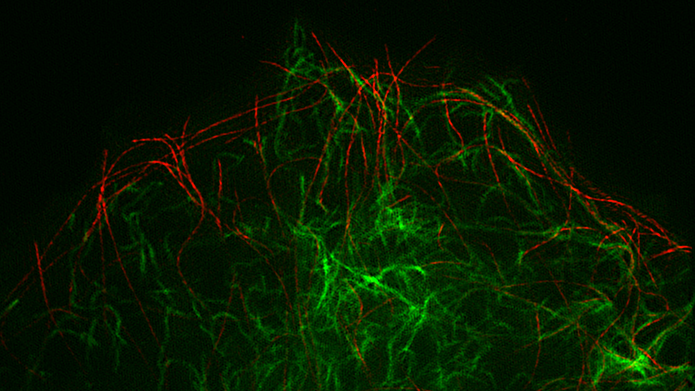
A human cell’s cytoskeleton – the protein network that supports its shape and function – is made of three components. Scientists know a lot about two of them, microfilaments and microtubules, but they’ve only recently had the technological advances to study the dynamics of the third in detail.

In a new pair of studies, Northwestern Medicine scientists help explain how this third component – slender, threadlike structures called intermediate filaments – moves and assembles to protect cells.

The work, led by Vladimir Gelfand, PhD, Leslie B. Arey Professor of Cell, Molecular, and Anatomical Sciences in the Department of Cell and Molecular Biology, may someday have applications for patients with genetic mutations that affect intermediate filaments and their important mechanical properties.

“Cells go through mechanical stress all the time. When we move, they essentially have to change their shape to resist the mechanical stress,” Gelfand said. “But intermediate filaments were not known as a very dynamic structure. So the big question was, how do they rearrange?”

In one paper, published in Molecular Biology of the Cell, Gelfand and first author Caroline Hookway, a doctoral student in the Northwestern University Interdepartmental Neuroscience Program (NUIN), showed that intermediate filaments are transported in cells along microtubules, one of the other components of the cytoskeleton.

The scientists were able to make this discovery using newly developed optical probes and superresolution microscopy at Northwestern University’s Nikon Imaging Center. Intermediate filaments are too small to visualize individually with traditional microscopy.

Gelfand authors-500
Vladimir Gelfand, PhD, Leslie B. Arey Professor of Cell, Molecular, and Anatomical Sciences in Cell and Molecular Biology (middle), with the first authors of the two papers, Caroline Hookway, an NUIN doctoral student (left), and Amélie Robert, PhD, a postdoctoral fellow (right).

They also found that intermediate filaments rearrange in a cell through an extensive severing and recombination process.

“Mature filaments get chopped up into bits and then those bits come back together to make a new filament,” Hookway said.

That process explains one way intermediate filaments are assembled. But they’re also formed from scratch – from a building block of 32 individual protein chains stacked together. In a second paper published in Proceedings of the National Academy of Sciences (PNAS), Gelfand and first author Amélie Robert, PhD, a postdoctoral fellow, demonstrated that pieces of this building block rapidly exchange subunits during the first step of intermediate filament assembly, in a way they can’t in fully developed filaments.

“You can readjust subunits of the building block in the beginning, but not once they’re all incorporated into the mature filament,” said Robert.

Both studies worked with an intermediate filament protein called vimentin, because it’s found in all connective tissue. In addition, epithelial cells start expressing this protein when they become cancer cells. For these reasons, findings involving vimentin could have broad applications.

In future research, the scientists hope to identify all the building block proteins in cells that interact with intermediate filaments and to find out their functions to understand how this cytoskeletal component is regulated.

Both published studies were supported by National Institutes of Health (NIH) National Institute of General Medical Science (NIGMS) grant P01GM09697, a program project grant led by Robert Goldman, PhD, chair of Cell and Molecular Biology. The study in PNAS was also supported by NIGMS grant R01 GM52111 and American Heart Association Fellowship 13POST16210010.

Cell and Developmental Biology Research
Share. Facebook Twitter Email

Related Posts

Genetic Variations Influence Drug Metabolism in Patients of African Descent

Feb 7, 2023

Potential Therapeutic Target for Schizophrenia Identified 

Feb 6, 2023

Longtime Alcohol Consumption Speeds Up Biological Aging 

Feb 3, 2023

Comments are closed.

Latest News

Workshop Teaches Medical Students Diagnosis Skills in Dermatology

Feb 7, 2023

Genetic Variations Influence Drug Metabolism in Patients of African Descent

Feb 7, 2023

Potential Therapeutic Target for Schizophrenia Identified 

Feb 6, 2023

Longtime Alcohol Consumption Speeds Up Biological Aging 

Feb 3, 2023

Grant Provides Imaging Technology to Department of Ophthalmology

Feb 2, 2023
  • News Center Home
  • Categories
  • Press Release
  • Media Coverage
  • Editor’s Picks
  • News Archives
  • About Us
Flickr Photos
Feinberg_In_Vivo_20221209_tcrawford-24
Feinberg_In_Vivo_20221209_tcrawford-16
Feinberg_In_Vivo_20221209_tcrawford-14
Feinberg_In_Vivo_20221209_tcrawford-5
Feinberg_In_Vivo_20221209_tcrawford-6
Feinberg_In_Vivo_20221209_tcrawford-10
Feinberg_In_Vivo_20221209_tcrawford-8
Feinberg_In_Vivo_20221209_tcrawford-18
Feinberg_In_Vivo_20221209_tcrawford
Feinberg_In_Vivo_20221209_tcrawford-23
Feinberg_In_Vivo_20221209_tcrawford-25
Feinberg_In_Vivo_20221209_tcrawford-26

Northwestern University logo

Northwestern University Feinberg School of Medicine

RSS Facebook Twitter LinkedIn Flickr YouTube Instagram
Copyright © 2023 Northwestern University
  • Contact Northwestern University
  • Disclaimer
  • Campus Emergency Information
  • Policy Statements

Type above and press Enter to search. Press Esc to cancel.