Feinberg
Northwestern Medicine | Northwestern University | Faculty Profiles

News Center

  • Categories
    • Campus News
    • Disease Discoveries
    • Clinical Breakthroughs
    • Education News
    • Scientific Advances
  • Press Release
  • Media Coverage
  • Podcasts
  • Editor’s Picks
    • COVID-19
    • Cardiology
    • Cancer
    • Neurology and Neuroscience
    • Aging and Longevity
    • Artificial Intelligence in Medicine
  • News Archives
  • About Us
    • Media Contact
    • Share Your News
    • News Feeds
    • Social Media
    • Contact Us
Menu
  • Categories
    • Campus News
    • Disease Discoveries
    • Clinical Breakthroughs
    • Education News
    • Scientific Advances
  • Press Release
  • Media Coverage
  • Podcasts
  • Editor’s Picks
    • COVID-19
    • Cardiology
    • Cancer
    • Neurology and Neuroscience
    • Aging and Longevity
    • Artificial Intelligence in Medicine
  • News Archives
  • About Us
    • Media Contact
    • Share Your News
    • News Feeds
    • Social Media
    • Contact Us
Home » Protein Marks Vital Component in Creation of Multi-Ciliated Cells
Scientific Advances

Protein Marks Vital Component in Creation of Multi-Ciliated Cells

By Roger AndersonOct 1, 2013
Share
Facebook Twitter Email
Cilia, in green, project outward from the cell and produce a wave-like motion that sweeps mucous away from the lungs, moves eggs from the ovary to the uterus, and drives cerebral spinal fluid through the ventricles of the brain. 

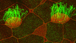
What started as a “random” dot under fluorescent microscopy more than five years ago is today the focal point of a paper in a major publication.

Discovered by Brian Mitchell, PhD, assistant professor of cell and molecular biology, the protein CCDC78 marks a significant advancement in understanding the formation of multi-ciliated cells.

Cilia, which resemble hair-like projections, are found in cells lining the trachea, where their wave-like motion sweeps mucous away from the lungs; in the fallopian tubes, where they are responsible for moving eggs from the ovary to the uterus; and in the brain, where they drive cerebral spinal fluid through the ventricles.

The body’s multi-ciliated cells represent an interesting biological variation in that they generate more than 100 centrioles, an organelle crucial for cell division that typically numbers just two. This increase in centrioles depends on the deuterosome, an uncharacterized structure thought to be the “factory” which churns them out.

Featured in the October 14 issue of Developmental Cell, Mitchell found that the dot he noticed a half-decade ago is actually the precise location of centriole amplification.

“Despite the many advances in molecular and cell biology over the past 50 years, somehow the deuterosome had remained molecularly uncharacterized,” Mitchell said. “Our work describes for the first time the mechanism that is required to generate hundreds of centrioles, which allow for the proper development of ciliated tissues.”

Brian Mitchell, PhD, assistant professor of cell and molecular biology, right, and postdoctoral fellow Deborah Klos-Dehring have identified a protein that leads to the development of multi-ciliated cells.

No matter the location, the multi-ciliated tissues of humans are remarkably similar and share many features with the multi-ciliated tissue found on the surface of developing Xenopus (frog) embryos.

Because of this, the animal model offers a “perfect” system for understanding the molecular processes required to form a functional multi-ciliated tissue.

Since centriole duplication is typically one of the first steps in the cell cycle and is critical to proper cell division, Mitchell’s lab remains interested in the creation of organelles in cells that make many cilia. Understanding how nature has uncoupled centriole duplication from the cell cycle could lead to insights into how the process goes awry during cancer development.

“Beginning the process of defining the mechanism allows us to then think about the difference between how these multi-ciliated cells make their centrioles versus how a dividing cell makes its centrioles,” said Mitchell, a member of the Robert H. Lurie Comprehensive Cancer Center of Northwestern University. “We now have the players and tools to dig deeper into the biochemistry of how these proteins are regulated.”

This research was supported by the National Institutes of Health through National Institute of General Medical Sciences grant R01GM089970, a pilot grant from the Northwestern University Skin Disease Research Center, an American Recovery and Reinvestment act grant, and a postdoctoral fellowship grant from the American Heart Association.

Cell and Developmental Biology Research
Share. Facebook Twitter Email

Related Posts

Humans are Not Just Big Mice: Identifying Science’s Muscle-Scaling Problem

Mar 20, 2023

Predicting Risk of Blood Clots in Brain Tumors

Mar 16, 2023

Understanding How Exercise Induces Systemic Metabolic Benefits

Mar 15, 2023

Comments are closed.

Latest News

Humans are Not Just Big Mice: Identifying Science’s Muscle-Scaling Problem

Mar 20, 2023

AOA Honors New Members

Mar 20, 2023

Celebrating Feinberg’s 2023 Match Day

Mar 17, 2023

Predicting Risk of Blood Clots in Brain Tumors

Mar 16, 2023

Understanding How Exercise Induces Systemic Metabolic Benefits

Mar 15, 2023
  • News Center Home
  • Categories
  • Press Release
  • Media Coverage
  • Editor’s Picks
  • News Archives
  • About Us
Flickr Photos
_5NM1245
230204_SERIO_MANDELL_Feinberg_Formal_0928
_5NM1715
_5NM0526
_5NM1026 (1)
_5NM1906
_5NM2173
230204_SERIO_MANDELL_Feinberg_Formal_0896
230204_SERIO_MANDELL_Feinberg_Formal_1113
230204_SERIO_MANDELL_Feinberg_Formal_1868
230204_SERIO_MANDELL_Feinberg_Formal_1237
230204_SERIO_MANDELL_Feinberg_Formal_1172

Northwestern University logo

Northwestern University Feinberg School of Medicine

RSS Facebook Twitter LinkedIn Flickr YouTube Instagram
Copyright © 2023 Northwestern University
  • Contact Northwestern University
  • Disclaimer
  • Campus Emergency Information
  • Policy Statements

Type above and press Enter to search. Press Esc to cancel.