Investigators from the laboratory of Derek Walsh, PhD, professor of Microbiology-Immunology, have discovered how human cytomegalovirus rewires intracellular mechanisms to control the movement of the cell nucleus, promoting infection and mediating cell migration, according to a recent study published in Proceedings of the National Academy of Sciences.
Human cytomegalovirus (HCMV) is a DNA virus that forms lifelong infection. In healthy individuals, the immune system prevents the virus from causing illness, but the virus can be harmful those who are immunocompromised and it is the leading cause of congenital birth defects, according to the Centers for Disease Control and Prevention.
During infection, HCMV replicates slowly over several days. During that time, the virus extensively remodels the infrastructure of healthy cells, including the nucleus. The precise mechanisms involved, however, have remained poorly understood, Walsh said.
“We have had a long-standing interest in understanding this cellular remodeling and in particular, how the virus exploits cytoskeletal interactions with the nucleus to control its shape and movement during infection. This is in part because a hallmark of HCMV infection is an expanded, distorted nucleus but until recently, precisely how and why this happens remained unclear,” Walsh said.
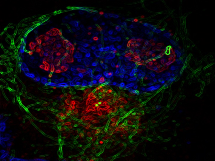
To better understand how HCMV affects nuclear movement, Walsh’s team used extended live cell imaging, or timelapse imaging, to study HCMV infection over several days. These viruses also expressed fluorescent proteins and fluorescently labeled cells, showing how the nucleus and cell changed shape over the course of infection.
Using a combination of live cell and fixed cell imaging, the investigators then observed how the nucleus moved during the later phase of infection as the virus drove cell migration.

Using these techniques, they discovered that HCMV encodes a kinase that disrupts lamin A/C organization.
Lamin A/C is a filament network, or mesh, that lines the inner nuclear membrane and organizes chromatin in the nucleus. Lamin A/C also interacts with the proteins SUN1 and SUN2, which form larger complexes called “LINCS” that extend into the cell’s cytoplasm and interact with cytoskeletal filaments, such as actin and microtubules, that position and control the nucleus during cell migration.
In addition to disrupting lamin A/C organization, the scientists discovered that HCMV also downregulates the SUN2 protein, which prevents this protein and others from interfering with the formation of microtubules that support nuclear movement and cell migration.
“This underscores the importance of microtubules in how the virus controls nuclear movement during infection,” Walsh said.
The findings demonstrate how HCMV exploits different cellular networks to control nuclear movement and shape, which is required for cell migration during infection. The findings may also inform the development of new therapeutic targets for both HCMV and other diseases, according to Walsh.
“While that might be some way down the road, this highlights the importance of basic research in identifying the underlying mechanisms of virus replication and what host factors they rely upon, which may form the basis for drug targets in the future,” Walsh said. “We’re now trying to understand the extent to which HCMV targets broader components of the lamin-based LINC complex system that control nuclear interactions with the cytoskeleton.”
Jamil Mahmud, PhD, a former postdoctoral fellow in the Walsh laboratory, and Ipsita Nandi, a postdoctoral fellow in the Walsh laboratory, were co-lead author of the study.
This work was supported by the National Institute of Allergy and Infectious Diseases grant R01AI141470.