Northwestern Medicine scientists have pioneered a new method to enhance bone regeneration therapies, as detailed in a study published in Nature Communications.
The new technique could revolutionize the design of orthopedic and craniofacial implants, offering a new strategy to enhance healing by harnessing the body’s own cellular machinery, said Guillermo Ameer, ScD, the Daniel Hale Williams Professor of Biomedical Engineering and Surgery, who was senior author of the study.
“Tissue damage because of trauma is a very common issue,” said Ameer, who leads the Querrey Simpson Institute for Regenerative Engineering. “To try and replace tissue, we sometimes use plastic or metals to try to fill in the defect that is typically formed when you lose tissue. What we’re trying to do with regenerative medicine is help restore that defect with natural tissue — basically your own tissue.”
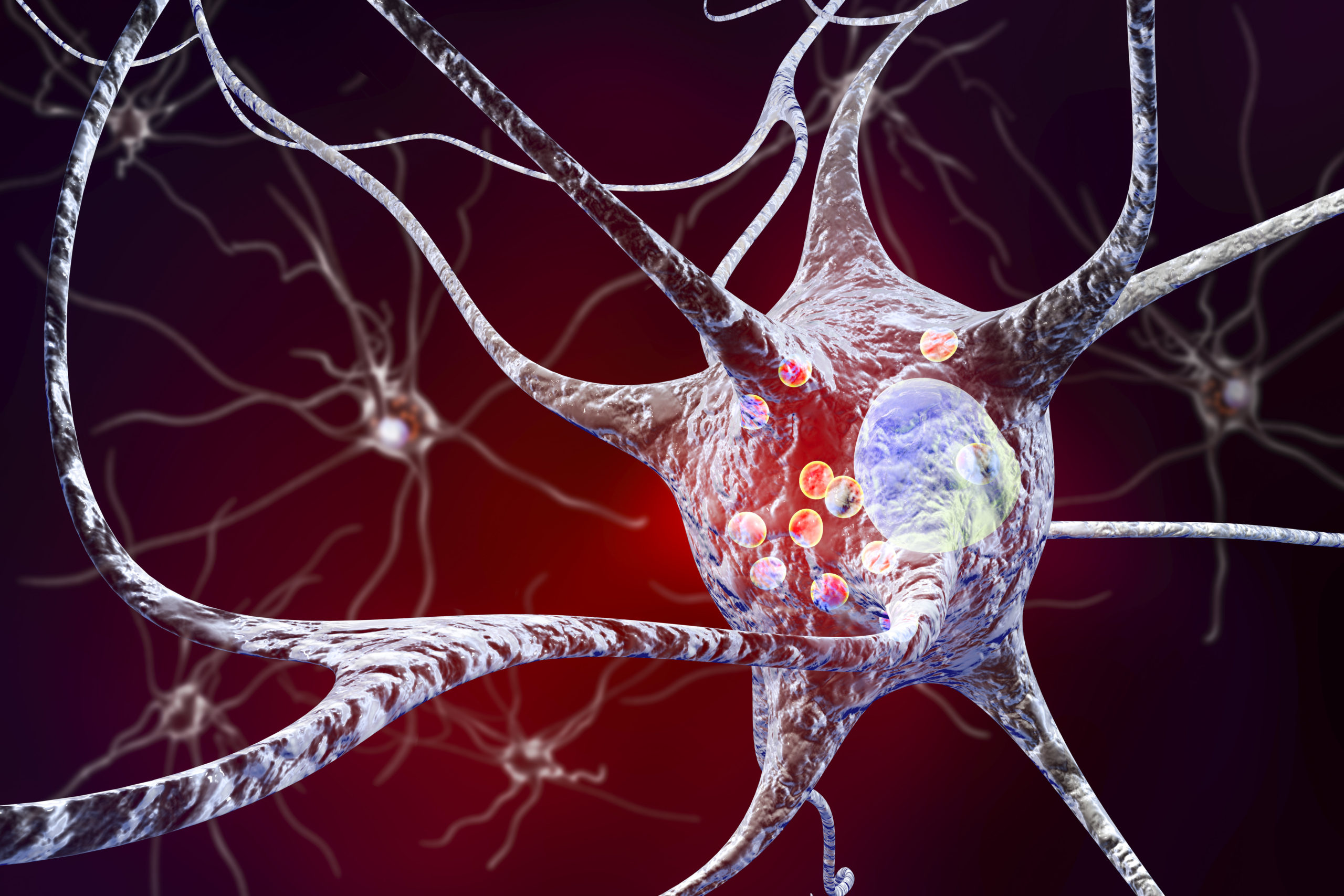
In previous work, investigators from the Ameer laboratory developed innovative implants with a unique engineered micropillar surface. These tiny pillars physically deform the nuclei of mesenchymal stem cells (MSCs) that attach to them. While previous studies have focused on how nuclear shape affects a cell’s internal fate, the new findings reveal that the altered cells also begin secreting proteins that enhance bone growth in neighboring cells.
In the current study, investigators analyzed fabricated implants during the process of bone growth. They found that when MSCs experience nuclear deformation due to the micropillars, they increase the secretion of proteins that organize the extracellular matrix —the structural network that supports tissues. This process promotes bone formation in nearby MSCs, even if those cells are not directly in contact with the implant.
To test the real-world potential of this discovery, the team implanted the micropillar devices into mice with cranial bone defects. They observed that the deformed-nucleus MSCs on the implants showed increased expression of Col1a2, a gene essential for collagen production and bone matrix formation. This led to enhanced bone regeneration in the defect area.
The findings highlight a phenomenon known as matricrine signaling, in which cells influence each other through changes in the extracellular matrix rather than direct contact or traditional signaling molecules. This opens new avenues for designing implants that not only support tissue structurally but also actively guide healing through cellular communication.
“When the nucleus of a cell is deformed by the micropillars, the environment and structure of its chromosomes change in such a way that production and secretion of proteins that tell cells to make bone is favored,” Ameer said. “Here, we identified a mechanism through which that process can promote new bone formation in nearby cells that are not in direct contact with the micropillars. It turns out that the secreted proteins modify and instruct the matrix that surrounds those nearby cells to support bone formation.”
Future studies may explore how this approach could be adapted for other tissues, such as cartilage, Ameer said.
“Cartilage loss is a big problem, especially in arthritis,” Ameer said. “The body can’t really regenerate it. But we have work that suggests that we can use a 3D printing method to help that process.”
The study was supported by the National Science Foundation Emerging Frontiers in Research and Innovation award no. 1830968, as well as grant EFMA-1830961. Additional funding was provided by the National Institutes of Health grants U54CA268084, R01CA228272 and R01DE030480.