Investigators led by Navdeep Chandel, PhD, the David W. Cugell, MD, Professor of Medicine in the Division of Pulmonary and Critical Care, have discovered how the metabolism of mitochondria supports T-cell proliferation and also prevents T-cell exhaustion in cancer and chronic infection, according to recent findings published in Nature Immunology.
The findings improve the understanding of how mitochondria support proper T-cell function and could inform the development of new targeted immunotherapies.
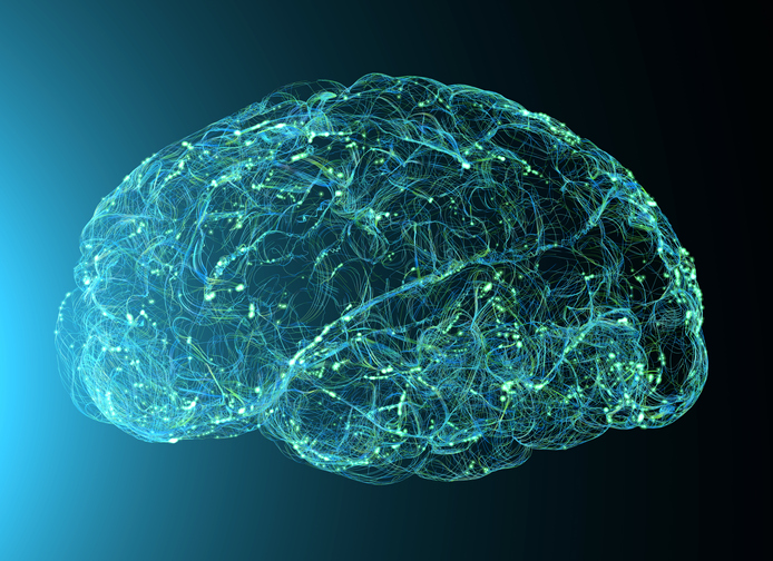
The mitochondrial electron transport chain (ETC) is a series of protein complexes inside mitochondria that facilitate the transfer of electrons and supports the generation of ATP, or energy for the cell.
Previous work from the Chandel laboratory had demonstrated mitochondrial ETC function was required for the proliferation of CD8+ T-cells, immune cells that help fight cancer and viral infection by recognizing their antigens. However, the precise mitochondrial ETC functions that promote CD8+ T-cell response and proliferation have remained unknown until now.
In the current study, the scientists developed mice lacking mitochondrial complex III, one of four complexes in the mitochondrial ETC that transports electrons and also delivers protons that support ATP generation and the production of reactive oxygen species (ROS) as cell signaling molecules.
From observing the deficient mice, the scientists discovered that impaired mitochondrial complex III function resulted in decreased cellular respiration and signaling molecules linked to ATP production. The loss of mitochondrial complex III in the CD8+ T-cells diminished their proliferation upon viral infection. Surprisingly, these cells also underwent rapid exhaustion upon acute antigen stimulation. Previous studies had only observed exhaustion under chronic antigen stimulation, according to the authors.
Additionally, they found that impaired mitochondrial complex III function reduced CD8+ T-cell memory formation, which helps the immune cells persist after initial infection and provide protection for subsequent infections.
“Every time you get a viral infection, you robustly respond to the virus and you clear it out, but you have a few memory T-cells that are sitting around that if you get the same virus again, they proliferate quickly to protect you,” said Chandel, who is also a professor of Biochemistry and Molecular Genetics, an investigator of the Chan Zuckerberg Biohub and a member of the Robert H. Lurie Comprehensive Cancer Center of Northwestern University.
To further investigate this, the scientists inserted C. intestinalis-derived alternative oxidase (AOX) protein in the mitochondrial complex III-deficient CD8+ T-cells; AOX can complement the loss of mitochondrial complex III without generating ROS. They found that while AOX prevented exhaustion as well as helped restore cell metabolism and proliferation, it did not restore memory formation, suggesting memory formation is linked to ROS generation.
“We saw memory precursor marker expression by those cells, showing that it’s not just an immediate death signal, but it’s a failure to form a terminal memory cell that can stick around,” said Elizabeth Steinert, PhD, research assistant professor of Medicine in the Division of Pulmonary and Critical Care and lead author of the study.
Overall, the findings demonstrate how mitochondrial respiration is required for T-cell proliferation and memory formation, which may help inform the development of new therapeutic strategies that target mitochondria in these cells, according to Chandel.
“This tells you that mitochondrial metabolism prevents exhaustion, mitochondrial metabolism supports proliferation and mitochondrial ROS is necessary to make memory. That really puts mitochondria at the center of T-cell biology, so maybe we should think about therapies that target mitochondria to rejuvenate them,” Chandel said.
Co-authors of the study include Beatriz Furtado Bruza, a graduate student in the Driskill Graduate Program in Life Sciences (DGP); Rogan Grant, PhD, a Schmidt Science Fellow at Northwestern; Karthik Vasan, a former student in the Medical Scientist Training Program (MSTP); Weiguo Cui, PhD, professor of Pathology in the Division of Experimental Pathology; and Samuel Weinberg, ’19 MD, ’19 PhD, assistant professor of Pathology in the Division of Experimental Pathology.
Chandel, Cui and Weinberg are also members of the Center for Human Immunobiology.
This work was supported by the National Institute of Health grants R01AI148190 and 5P01HL154998, the Cancer Research Institute, and by Schmidt Science Fellows in partnership with the Rhodes Trust.