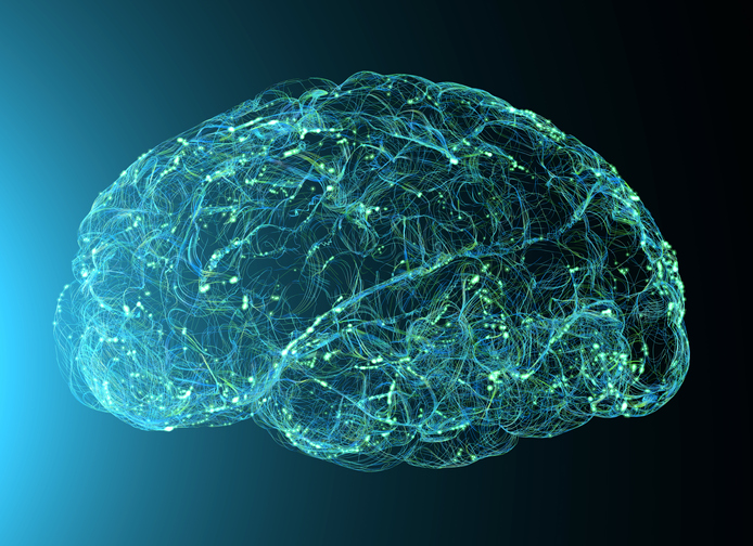
An artificial intelligence-based imaging approach may be an effective tool for distinguishing patients with Parkinson’s disease from those with other closely related diseases sooner than current methods, according to a recent study published in JAMA Neurology.
Previous work suggests that advanced MRI imaging paired with artificial intelligence, specifically machine learning, may be an effective approach for differentiating patients with Parkinson disease (PD), multiple system atrophy, parkinsonian variant (MSA-P) and progressive supranuclear palsy (PSP).
These diseases share similar symptoms, including slowness of movement, muscle stiffness and tremor, making it difficult for clinicians to clearly differentiate one condition from the other. This underscores the need for better diagnostic approaches that can help clinicians make accurate diagnoses earlier, said Rizwan Akhtar, MD, PhD, assistant professor in the Ken and Ruth Davee Department of Neurology’s Division of Movement Disorders and a co-author of the study.
“It can be hard to confirm a diagnosis of MSA-P or PSP early on in the disease. The most useful thing at the moment is to follow patients over time and see how they change, what is the pace of their progression, and whether they have any unusual features that could steer us away from Parkinson’s disease, and this process can take some time,” Akhtar said. “If a person has one of these atypical disorders, we want to identify that as soon as we can because we need to counsel the family and counsel the patient about what to expect. We may want to recruit them into other clinical trials that are testing interventions and the earlier you start those interventions, the better.”
In the current study, advanced brain MRI scans from 249 patients (99 patients with PD, 53 with MSA-P, and 97 with PSP) and a retrospective cohort of 396 patients (211 patients with PD, 98 with MSA and 87 with PSP) enrolled at 21 study sites across the U.S. and Canada were collected.
The scans were then used to train a machine learning algorithm to distinguish subtle differences in the imaging to distinguish patients with PD versus atypical parkinsonism, MSA versus PSP, PD versus MSA, and PD versus PSP. The clinical diagnoses were validated by the investigators.
Overall, the investigators found the AI-based imaging approach could distinguish PD from atypical parkinsonism with 96 percent sensitivity and PD from MSA and PSP with 98 percent sensitivity. The approach also predicted postmortem neuropathology in approximately 94 percent of autopsy cases, whereas clinical diagnosis was confirmed in only 81.6 percent of cases.
“That was remarkable because for most biomarkers, once they reach about 90 percent sensitivity, we think that’s pretty good, and this was far more than that,” Akhtar said.
The findings suggest that clinicians could use the MRI imaging to diagnose these uncommon neurodegenerative forms of parkinsonism sooner than is currently possible.
“I think the long-term goal is to develop this approach into a tool that can help diagnosis patients sooner to try and get them the help they need as early as possible. In my own work, I hope to apply these types of methods in our aging studies at the Mesulam Center for Cognitive Neurology and Alzheimer’s Disease,” said Todd Parrish, PhD, professor of Radiology and a professor of Biomedical Engineering at the McCormick School of Engineering, who was also a co-author of the study.
The approach may also help shorten the time it takes for clinicians to arrive at a conclusive diagnosis, a current issue in the field, according to Akhtar.
“One of the major issues in our field is that access to specialists who can clinically distinguish between these three illnesses is limited,” Akhtar said. “The reality is it’s not easy for every patient to come to a tertiary care center, whereas an MRI like this one can be done anywhere. One of the goals of the study is to try to make the algorithm available online for a larger audience. Ideally a person can get an MRI locally and have it uploaded and analyzed by the algorithm.”
This work was supported by the National Institutes of Health grant number U01NS119562.