Northwestern Medicine scientists have shed new light on the inner workings of some of the smallest cellular structures, according to a study published in the Journal of Cell Biology.
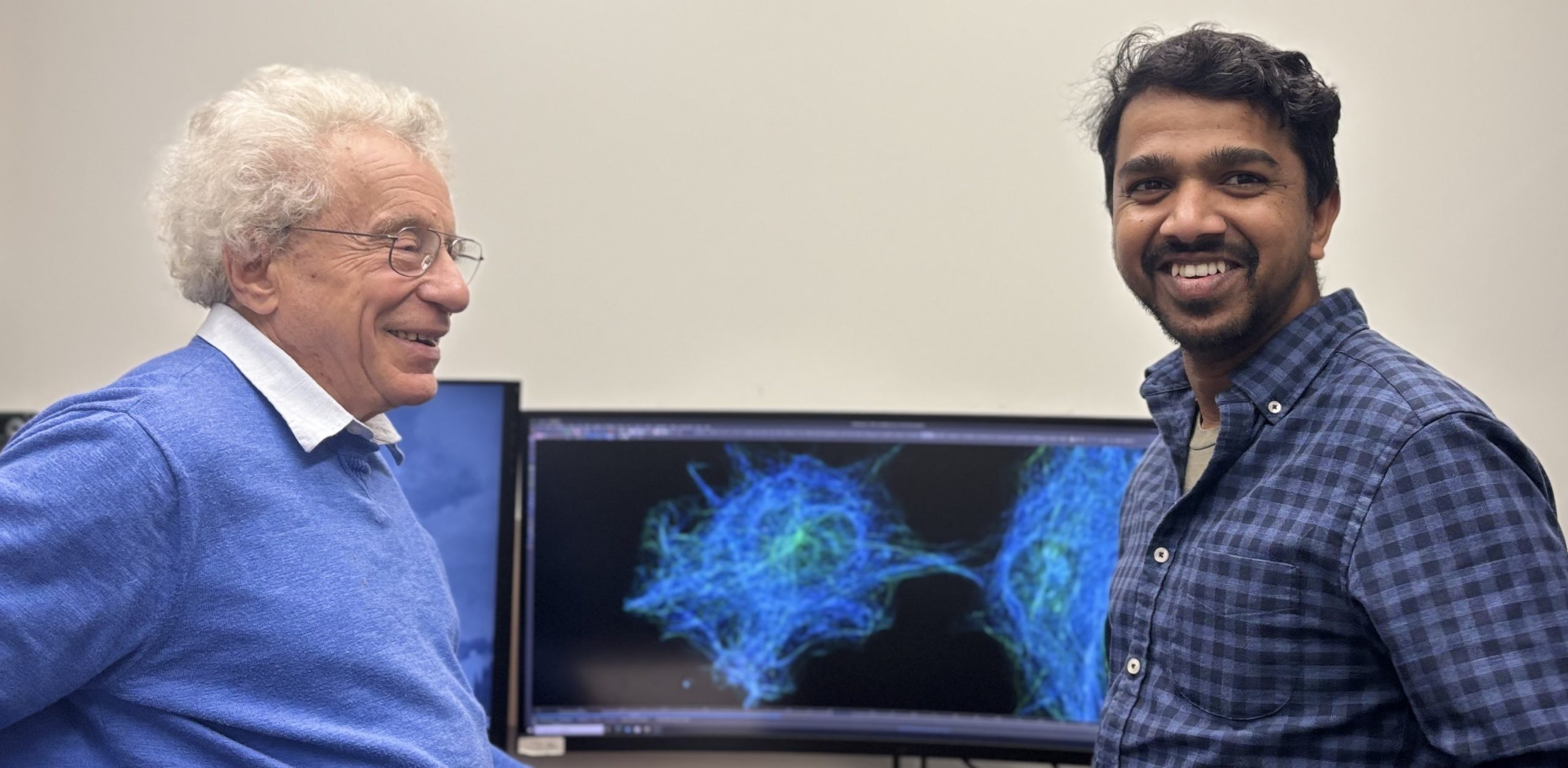
Vimentin, a protein that forms structures called intermediate filaments, is an important component of the cellular cytoskeleton that acts as a kind of load-bearing scaffolding and is critical in helping cells keep their shape and move. Advancements in laboratory technology have only recently made it possible to observe the dynamics of intermediate filaments within a single cell, said Vladimir Gelfand, PhD, the Leslie B. Arey Professor of Cell, Molecular, and Anatomical Sciences and senior author of the study.

“Essentially, these filaments are typically considered as the most non-dynamic component of the cytoskeleton,” said Gelfand, who is a member of the Robert H. Lurie Comprehensive Cancer Center of Northwestern University. “People generally believe that filaments just help cells to keep their shape and prevent mechanical damage. But a long time ago, we started to suspect that the filaments are more dynamic than people think.”
In the present study, investigators in the Gelfand laboratory developed a novel technique to observe vimentin filaments within a living single cell. By employing single-particle tracking and volume electron microscopy reconstruction, investigators were able to track individual vimentin filaments.
They observed that vimentin filaments actively move in a cell along microtubules, cellular “highways” that facilitate the transfer of cellular cargo. The filaments could be seen moving individually to interact with neighboring microtubules, contrary to previous assumptions that vimentin filaments acted as part of tightly-linked bundles.
“In a joint work with the group of Jennifer Lippincott-Schwartz and her group at HHMI Janelia Research Campus in Virginia, we showed that intermediate filaments are not bundled. They are individual filaments,” said Bhuvanasundar Ranganathan, PhD, a research associate in the Gelfand laboratory and first author of the study. “We showed that they are dynamic in every part of the cell.”
Taken together, the findings demonstrate for the first time that vimentin filaments are a complex part of the cellular cytoskeleton, Ranganathan said.
“Vimentin filaments play a major role in cell shape and cell motility,” Ranganathan said. “Further, the technique we developed will be helpful for studying how it functions in cell migration.”
In the future, Gelfand and Ranganathan will study how vimentin filaments contribute to the movement of organelles within the cell.
“We plan to study its function in normal cells and extrapolate how it might contribute to disease, such as cancer,” Ranganathan said.
The study was supported by grant 2R35-GM131752 from the National Institute of General Medical Sciences.