
A Northwestern Medicine study has uncovered novel cellular mechanisms within the retina, findings that could help advance the development of targeted therapeutics for diseases and conditions impacting vision, according to findings published in Nature Communications.
Cone synapses inside the eye’s retina help the brain process changes in light. It’s a unique synapse because it is has evolved to signal changes in light intensity, said Steven DeVries, MD, PhD, the David Shoch, MD, PhD, Professor of Ophthalmology and senior author of the study.
“Counterintuitively, cone neurotransmitter release is high in the dark and reduced by light. When the light’s brighter, the reduction is larger. When the lights are dimmer, it’s smaller; it operates differently from most synapses which use an increase in transmitter release to signal all-or-nothing, digital action potentials,” DeVries said.
Unlike most other synapses in the brain, each individual cone synapse is connected to more than a dozen different types of post-synaptic neurons, the bipolar cells, which relay information in parallel to the inner retina. In the inner retina, these parallel streams not only contribute to conscious vision but also to subconscious processes like gaze stabilization.
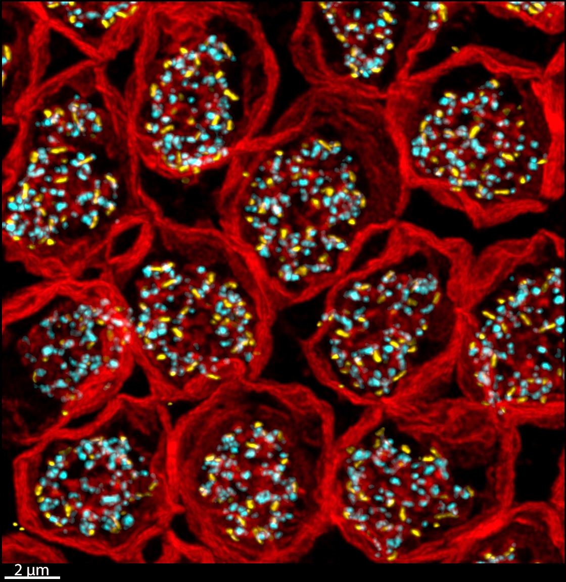
In the current study, which involved non-human mammalian retinas, the investigators first used super-resolution microscopy to map out the locations of transmitter release sites, transmitter re-uptake proteins and post-synaptic contacts at the cone synapse. Then they used an approach called “synaptic accounting,” to relate the amount of transmitter released by a cone to the responses in each post-synaptic bipolar cell type.
“Transmitter is released in packets or quanta when a vesicle fuses with the presynaptic membrane. Since most synapses involve one-to-one direct contacts across a narrow cleft, it is assumed that one detected quantum equals one released quantum. The cone synapse has a different design that voids this assumption. We developed a way to stimulate a cone and count the vesicles that are released while at the same time counting the number of vesicles that are detected by the postsynaptic neuron,” DeVries said.
Using these techniques, the investigators showed that certain bipolar cell types respond to individual fusion events and total quanta while other types respond to degrees of locally coincident events, creating a nonlinear summation. These differences are caused by a combination of factors specific to each bipolar cell type, including diffusion distance, contact number, receptor affinity, and proximity to transporters.
“The outer retina uses the same toolbox as elsewhere in the central nervous system, like vesicles, synaptic release zones and postsynaptic receptors, but organizes these elements in novel ways to accomplish a different, very localized type of processing. Analog processing is also found in the dendritic tree of central nervous system neurons, where the bulk of calculation, both linear and nonlinear, occurs,” DeVries said.
According to DeVries, one next step for his team includes using a new, more powerful type of super-resolution microscopy to determine the protein components that make up cone synapses.
“One of the ways that the different bipolar cells divide the cone signal up is that some of them are very sensitive to small signals and others require strong signals to respond; the ‘strong signal’ or high threshold bipolar cell has a unique type of insensitive post-synaptic receptor. We would also like to identify this receptor,” DeVries said.
This work was supported by the National Institutes of Health grant R01 EY012141, an unrestricted grant to the Dept. of Ophthalmology from Research to Prevent Blindness, an International Travel Award from RPB, and JSPS KAKENHI grant #19H01140.