Feinberg
Northwestern Medicine | Northwestern University | Faculty Profiles

News Center

  • Categories
    • Campus News
    • Disease Discoveries
    • Clinical Breakthroughs
    • Education News
    • Scientific Advances
  • Press Releases
  • Media Coverage
  • Podcasts
  • Editor’s Picks
    • COVID-19
    • Cardiology
    • Cancer
    • Neurology and Neuroscience
    • Aging and Longevity
    • Artificial Intelligence in Medicine
  • News Archives
  • About Us
    • Media Contact
    • Share Your News
    • News Feeds
    • Social Media
    • Contact Us
Menu
  • Categories
    • Campus News
    • Disease Discoveries
    • Clinical Breakthroughs
    • Education News
    • Scientific Advances
  • Press Releases
  • Media Coverage
  • Podcasts
  • Editor’s Picks
    • COVID-19
    • Cardiology
    • Cancer
    • Neurology and Neuroscience
    • Aging and Longevity
    • Artificial Intelligence in Medicine
  • News Archives
  • About Us
    • Media Contact
    • Share Your News
    • News Feeds
    • Social Media
    • Contact Us
Home » Map of Broken Brain Networks Shows Why People Lose Speech in Language-Based Dementia
Disease Discoveries

Map of Broken Brain Networks Shows Why People Lose Speech in Language-Based Dementia

By Kristin SamuelsonSep 13, 2019
Share
Facebook Twitter Email


Illustration of the three brain regions associated with speech production and the networks between them that, when broken, lead to speech impairments. Solid orange lines depict significantly lower connectivity between brain regions (in red) in PPA. IFG = Inferior Frontal Gyrus; MTG = Middle Temporal Gyrus; ATL = Anterior Temporal Lobe. PPA = Primary Progressive Aphasia; PPA-G = Nonfluent/agrammatic variant of PPA; PPA-L = Logopenic variant of PPA; PPA-S = Semantic variant of PPA.

‘Now we know where to target peoples’ brains to attempt to improve their speech’

Borna Bonakdarpour, MD, assistant professor of Neurology in the Division of Behavioral Neurology and member of the Mesulam Center for Cognitive Neurology and Alzheimer’s Disease, was the lead author of the study.

For the first time, Northwestern Medicine scientists have pinpointed the location of dysfunctional brain networks that lead to impaired sentence production and word finding in primary progressive aphasia (PPA), a form of dementia in which patients often lose their language rather than their memory or thought process.

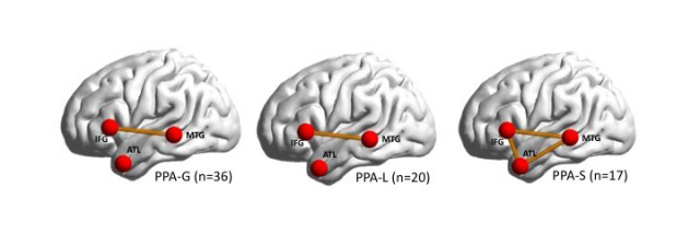
With this discovery, published in the journal Cortex, the scientists have drawn a map that illustrates three regions in the brain that fail to talk to each other, inhibiting a person’s speech production, word finding and word comprehension. For example, some people can’t connect words to form sentences, others can’t name objects or understand single words like “cow” or “table.”

The map can be used to target those brain regions with therapies, such as transcranial magnetic stimulation (TMS), to potentially improve an affected person’s speech.

“Now we know where to target people’s brains to attempt to improve their speech,” said lead author Borna Bonakdarpour, MD, assistant professor of in the Ken and Ruth Davee Department of Neurology in the Division of Behavioral Neurology, a member of the Mesulam Center for Cognitive Neurology and Alzheimer’s Disease as well as a Northwestern Medicine neurologist.

PPA occurs in patients with neurodegenerative disorders, including Alzheimer’s disease and frontotemporal degeneration.

Interactions among three main regions in the brain is responsible for how people process words and sentences. PPA occurs when there is a lack of connectivity among these areas. Different patterns of connectivity failure among these regions can cause different subtypes of PPA.

The findings were published Sept. 1 in the journal Cortex. The large study (73 patients) recruited from the extensive pool of patients with PPA at Northwestern’s Cognitive Neurology and Alzheimer Disease Center, one of the largest centers in the world.

Previous research used structural MRI to locate only regions of the brain that had atrophied, and scientists did not clearly know how physiological impairment in these regions correlated with symptoms a patient was experiencing. This study is novel because it examined brain regions that were still functional (had not atrophied) and focused on the networks among the functional regions to see if they were connecting or not. This allowed the scientists to correlate the functional areas in the brain with symptoms of patients with PPA.

“Previous studies of structural changes in the brain were like archeology, in which scientists were locating areas of the brain that had already died,” Bonakdarpour said. “But we are looking at the parts of the brain that are still alive, which makes them much easier to target with treatment.”

Bonakdarpour and his colleagues have begun testing TMS on the three targeted brain regions in healthy individuals with the goal of applying it to patients with PPA in a future clinical trial.

Read more about the subtypes of PPA on the Cognitive Neurology and Alzheimer’s Disease Center website.

Neurology and Neuroscience Research
Share. Facebook Twitter Email

Related Posts

Mar 29, 2023

Adolescent Sexual Health Program Receives Funding for Social Marketing Campaign

Mar 29, 2023

The Future of IgE-Mediated Allergy Research and Treatments

Mar 29, 2023

Investigating Protein’s Role in Hearing Loss

Mar 27, 2023

Comments are closed.

Latest News

Adolescent Sexual Health Program Receives Funding for Social Marketing Campaign

Mar 29, 2023

The Future of IgE-Mediated Allergy Research and Treatments

Mar 29, 2023

Weintraub Appointed to Illinois Supreme Court Commission on Elder Law

Mar 28, 2023

Investigating Protein’s Role in Hearing Loss

Mar 27, 2023

Sex-Specific Mechanisms for Major Depressive Disorder Identified in Response to Dysregulated Stress Hormones

Mar 23, 2023
  • News Center Home
  • Categories
  • Press Release
  • Media Coverage
  • Editor’s Picks
  • News Archives
  • About Us
Flickr Photos
20230317_NM651
20230317_NM610
20230317_NM569
20230317_NM537
20230317_NM331
20230317_NM323
20230317_NM316
20230317_NM336
20230317_NM626
20230317_NM662
20230317_NM655
20230317_NM642

Northwestern University logo

Northwestern University Feinberg School of Medicine

RSS Facebook Twitter LinkedIn Flickr YouTube Instagram
Copyright © 2023 Northwestern University
  • Contact Northwestern University
  • Disclaimer
  • Campus Emergency Information
  • Policy Statements

Type above and press Enter to search. Press Esc to cancel.