Northwestern Medicine scientists have identified a component of the herpesvirus that “hijacks” machinery inside human cells, allowing the virus to rapidly and successfully invade the nervous system upon initial exposure.
Led by Gregory Smith, PhD, associate professor in microbiology and immunology, investigators found that viral protein 1-2, or VP1/2, allows the herpesvirus to interact with cellular motors, known as dynein. Once the protein has overtaken this motor, the virus can speed along intercellular highways, or microtubules, to move unobstructed from the tips of nerves in skin to the nuclei of neurons within the nervous system.
This is the first time researchers have shown a viral protein directly engaging and subverting the cellular motor; most other viruses passively hitch a ride into the nervous system.
“This protein not only grabs the wheel, it steps on the gas,” says Smith. “Overtaking the cellular motor to invade the nervous system is a complicated accomplishment that most viruses are incapable of achieving. Yet the herpesvirus uses one protein, no others required, to transport its genetic information over long distances without stopping.”
Herpesvirus is widespread in humans and affects more than 90 percent of adults in the United States. It is associated with several types of recurring diseases, including cold sores, genital herpes, chicken pox, and shingles. The virus can live dormant in humans for a lifetime, and most infected people do not know they are disease carriers. The virus can occasionally turn deadly, resulting in encephalitis in some.
Until now, scientists knew that the herpesvirus travels quickly to reach neurons located deep inside the body, but the mechanism by which they advance remained a mystery.
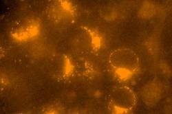
Smith’s team conducted a variety of experiments with VP1/2 to demonstrate its important role in transporting the virus, including artificial activation and genetic mutation of the protein. The team studied the herpesvirus in animals, and also in human and animal cells in culture under high-resolution microscopy. In one experiment, scientists mutated the virus with a slower form of the protein dyed red, and raced it against a healthy virus dyed green. They observed that the healthy virus outran the mutated version down nerves to the neuron body to insert DNA and establish infection.
“Remarkably, this viral protein can be artificially activated, and in these conditions it zips around within cells in the absence of any virus. It is striking to watch,” Smith says.
He says that understanding how the viruses move within people, especially from the skin to the nervous system, can help better prevent the virus from spreading.
Additionally, Smith says, “By learning how the virus infects our nervous system, we can mimic this process to treat unrelated neurologic diseases. Even now, laboratories are working on how to use herpesviruses to deliver genes into the nervous system and kill cancer cells.”
Smith’s team will next work to better understand how the protein functions. He notes that many researchers use viruses to learn how neurons are connected to the brain.
“Some of our mutants will advance brain mapping studies by resolving these connections more clearly than was previously possible,” he says.
This work was funded by NIH grant R01 AI056346, R01 EY017809, and by the training program in Immunology and Pathogenesis from the NIH (T32AI07476). It was published in the journal Cell Host & Microbe.


