This story was originally published in the December 2025 issue of the Breakthroughs newsletter.
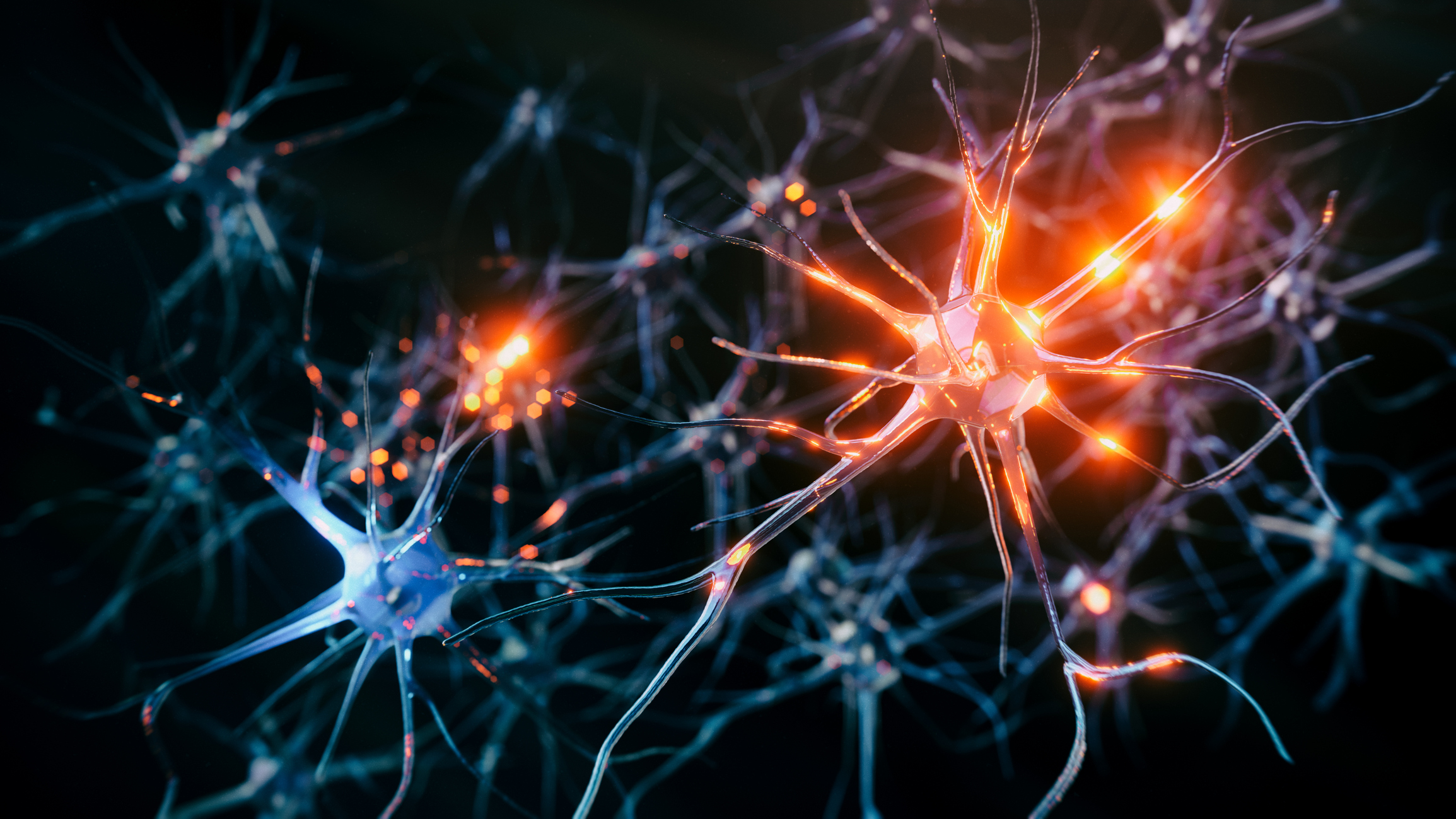
The human brain is often described as the most complex network in existence, with billions of neurons exchanging signals across intricate pathways. Understanding how the brain’s communication networks operate is key to discovering insights into cognition, behavior and neurological diseases. Recent research led by Feinberg investigators is revealing how different regions of the brain coordinate and process information, and how disruptions in these networks can impact health.
By analyzing these dynamic systems, scientists are finding new ways to understand the complexity of the brain to inform future diagnostics and treatment. In this story, we round up recent studies from Feinberg investigators that provide insight into how the brain functions, controls language and ‘forgets’ what is no longer needed.
Shedding Light on Neuronal Mechanisms
Investigators led by D. James Surmeier, PhD, the Nathan Smith Davis Professor and chair of Neuroscience, have uncovered a new way in which neurons in the brain “forget” associations that help guide behavior and habits, according to a study published in Cell Reports.
In addition to shedding light on basic brain mechanisms, the findings could also prove useful in treating Parkinson’s disease, said Surmeier, who was senior author of the study.
In the study, Surmeier and a team of Northwestern Medicine scientists set out to understand how spiny projection neurons — the principal neurons in the striatum, a key part of the brain circuitry controlling decision-making — are affected by neuronal plasticity, which is critical to the brain’s ability to change and adapt over time in response to life experiences.
“Some years ago, we discovered a novel form of long-term synaptic depression in spiny projection neurons that was triggered by a gaseous messenger called nitric oxide,” Surmeier said. “The stratum is one of the few places in the brain that has very high levels of the signaling molecules that respond to nitric oxide. We wanted to get a better understanding of the role that form of plasticity played in controlling behavior.”
Moving forward, Surmeier and his collaborators will continue to study the mechanisms of neuronal plasticity using cutting-edge techniques, he said.
“One of the things that we’re particularly excited about is that the tools we have to monitor and manipulate brain circuits has rapidly expanded, deepening our understanding of how Parkinson’s disease affects the brain and giving us strategies for reversing the changes in circuitry that cause symptoms,” he said. “We also are excited about new tools to manipulate nitric oxide signaling being developed by the Silverman lab here at NU.”
Utilizing Imaging to Understand Social Experiences
Another study published in Science Advances, led by Rodrigo Braga, PhD, assistant professor in the Ken and Ruth Davee Department of Neurology‘s Division of Epilepsy and Clinical Neurophysiology, and his team, sought to better understand how humans evolved to become so skilled at thinking about what’s happening in other peoples’ minds. The findings could have implications for one day treating psychiatric conditions such as anxiety and depression.

“We spend a lot of time wondering, ‘What is that person feeling, thinking? Did I say something to upset them?’” said Braga, who was senior author of the study. “The parts of the brain that allow us to do this are in regions of the human brain that have expanded recently in our evolution, and that implies that it’s a recently developed process. In essence, you’re putting yourself in someone else’s mind and making inferences about what that person is thinking when you cannot really know.”
The study found the more recently evolved and advanced parts of the human brain that support social interactions — called the social cognitive network — are connected to and in constant communication with an ancient part of the brain called the amygdala.
Within the amygdala, there’s a specific part called the medial nucleus that is very important for social behaviors. This study was the first to show the amygdala’s medial nucleus is connected to newly evolved social cognitive network regions, which are involved in thinking about other people. This link to the amygdala helps shape the function of the social cognitive network by giving it access to the amygdala’s role in processing emotionally important content.
Both anxiety and depression involve amygdala hyperactivity, which can contribute to excessive emotional responses and impaired emotional regulation, according to Donnisa Edmonds, first author of the study and PhD student in the Braga lab. Currently, someone with either condition could receive deep brain stimulation for treatment, but since the amygdala is located deep within the brain, directly behind the eyes, it means having an invasive, surgical procedure.
“Through this knowledge that the amygdala is connected to other brain regions — potentially some that are closer to the skull, which is an easier region to target — that means people who do transcranial magnetic stimulation (TMS) could target the amygdala instead by targeting these other regions,” Edmonds said.
Identifying Critical Language Connections
A third study published in Nature Communications may better inform doctors about which areas of the brain to preserve prior to surgery. The study expands the understanding of how language is encoded in the brain and identifies key features of critical sites in the cerebral cortex that work together to produce language.
Mark Slutzky, ‘02 MD, ‘00 PhD, ‘06 GME, professor of Neurology in the Division of Comprehensive Neurology, was the senior author. He and his team identified the critical language connector sites by recording electrical signals from the cortex of the brain in patients with epilepsy or brain tumors while the patients read words aloud. Investigators then analyzed the signals using graph theory methods and machine learning to predict which sites in the network were critical.

“This discovery could help us be more precise and efficient when we’re mapping language sites before surgery,” Slutzky said. “It could help us augment the way surgeons do this mapping, so ultimately it could potentially shorten the time needed for stimulation or possibly eliminate the stimulation and just record electrical signals.”
People with brain tumors or epilepsy who need surgery often undergo functional mapping using direct electrical stimulation of the brain to try to identify critical parts of the brain (particularly in the cerebral cortex) so neurosurgeons know which sites to avoid removing to preserve language.
Currently, many patients with brain tumors undergo between 20 and 60 minutes of stimulation while they are awake in surgery. The technique is not perfect for identifying the language sites: results can be false-negative or false-positive, and the process can cause seizures.
Scientists recorded electrical signals from the surface of the cortex in 16 patients (at Northwestern Memorial Hospital and at Johns Hopkins Hospital) with either epilepsy or brain tumors. The electrode arrays were either implanted in people with epilepsy as part of their seizure monitoring prior to surgery or placed on the brain temporarily in the operating room, while patients with tumors underwent awake brain surgery and mapping.
“When someone is speaking, many sites in the brain are active, yet only a handful of those sites are identified as critical to those functions by being perturbed during stimulation,” Slutsky said. “Answering this question could help us understand how electrical stimulation affects the brain and how the brain produces spoken language.”
As scientists continue to map the neuronal networks in the brain, Feinberg investigators are discovering new insights that enhance what we understand about the brain and how this information can be used to improve treatment for cognitive disorders. This work is critical to understanding the brain’s networks and a vital step toward improving human health and quality of life.
Olivia Dimmer and Kristin Samuelson contributed to this story.