Northwestern Medicine investigators have developed a new imaging approach to more accurately assess blood flow in the spinal cord, a method that could be used to better inform treatment for neurological diseases and injuries, as described in a recent study published in Scientific Reports.
“We’ve taken MRI methods that we’ve firmly established in the brain, and with a lot of attention to detail and many small improvements, we’ve made this critical jump to use them in the spinal cord, and I find that pretty exciting,” said Molly Bright, DPhil, assistant professor of Physical Therapy and Human Movement Sciences and of Biomedical Engineering in the McCormick School of Engineering, who was senior author of the study.
Impaired vascular function in the spinal cord contributes to many neurological diseases, including traumatic spinal cord injury and degenerative cervical myelopathy — a condition where age-related damage to the spinal discs compresses the spinal cord, which can cause progressive weakness, numbness and difficulty with coordination.
Measuring changes in blood supply to spinal cord tissue is essential for guiding preventive care measures and treatment. However, existing methods have yet to measure these changes accurately, Bright said.
To address this gap, Bright and her team developed a functional MRI (fMRI)-based approach to map spinal cord vascular reactivity, or how well blood vessels can dilate to increase blood flow to the spinal cord tissue. FMRI is most commonly used to map neural activity by measuring changes in blood flow and, therefore, an excellent imaging tool for studying blood supply directly, Bright said.
“For most of my career I’ve used fMRI not to look at just neural activity, but also to look at the plumbing, and how the vascular system is supporting the nervous system. This is important for many neurodegenerative diseases because neural and vascular health are so interconnected,” Bright said.
To validate their approach, study participants underwent a series of fMRI scans. During the scans, participants were asked to repeatedly hold their breath for short periods, which causes carbon dioxide to build up and triggers blood vessels to dilate, increasing blood flow.
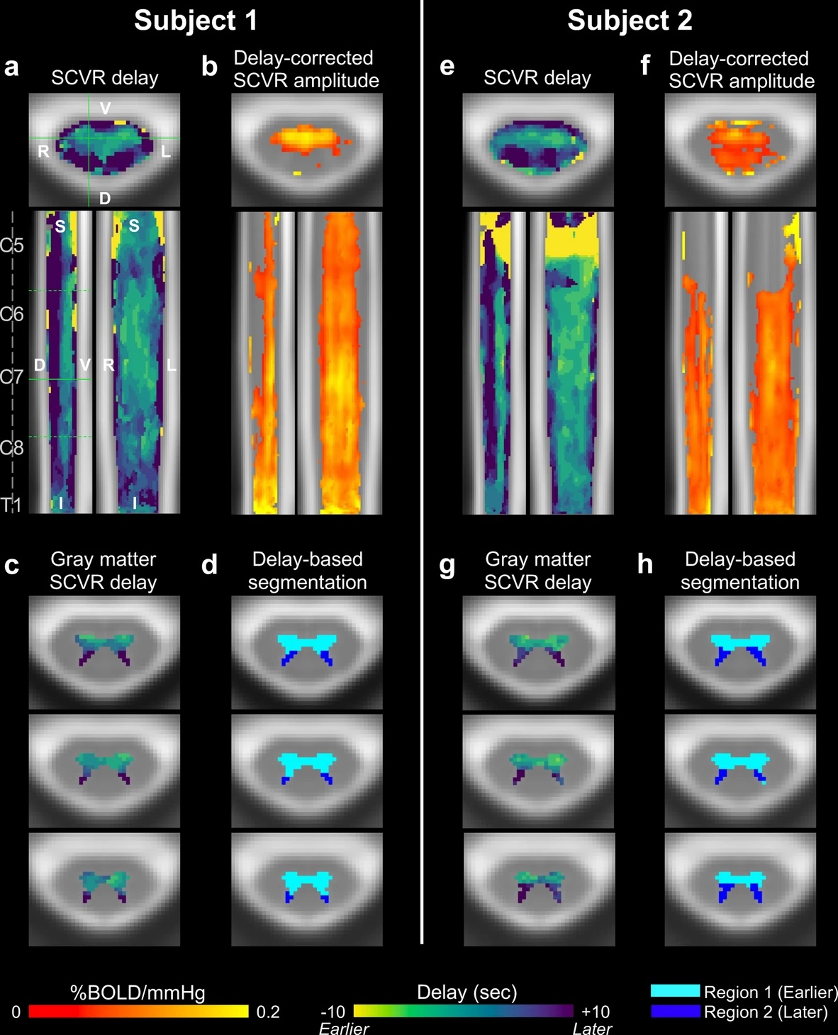
From these imaging scans, the scientists were then able to create maps of vascular function in the spinal cord, which revealed that certain regions of the spinal cord responded at different times.
“This was actually very consistent across people,” Bright said. “We didn’t really know to look for this ahead of time, but it might be that we’re seeing the circulation patterns of different arteries that supply blood to the spinal cord, which I don’t think have been mapped before.”
Overall, the approach can provide more detailed information about spinal cord vascular health, which can help guide treatment strategies for spinal cord injuries and diseases, Bright said.
“For spinal cord injury, we might be able to use this type of vascular mapping as well as some of the more traditional fMRI neural mapping to understand whether interventions are improving function in a damaged part of the cord,” Bright said.
Bright added that the approach could also help improve preventive care for patients with degenerative cervical myelopathy.
“If we can detect that the vascular supply is impaired in the area of cord compression in certain older adults with degenerative disc disease, then we can potentially identify who needs more monitoring or early intervention,” Bright said.
Co-authors of the study include Milap Sandhu, PhD, assistant professor of Physical Medicine and Rehabilitation, and Todd Parrish, PhD, professor of Radiology and of Biomedical Engineering at the McCormick School of Engineering.
Kimberly Hemmerling, PhD, a former graduate student in Northwestern’s Biomedical Engineering program, was the lead author of the study.
This work was supported by the Center for Translational Imaging at Northwestern University and the Craig H. Neilsen Foundation grant 595499.